File:Astrocyte5.jpg
From WikiMD's WELLNESSPEDIA

Size of this preview: 800 × 588 pixels. Other resolutions: 320 × 235 pixels | 640 × 470 pixels | 1,024 × 753 pixels | 1,309 × 962 pixels.
Original file (1,309 × 962 pixels, file size: 1.75 MB, MIME type: image/jpeg)
Summary
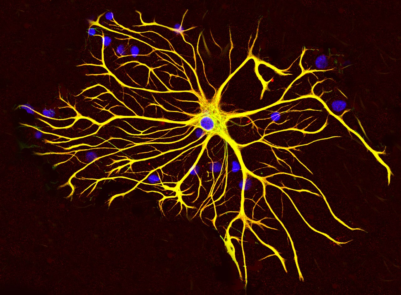
| Description |
English: An astrocyte cell grown in tissue culture stained with antibodies to GFAP and vimentin. The GFAP is coupled to a red fluorescent dye and the vimentin is coupled to a green fluorescent dye. Both proteins are present in large amounts in the intermediate filaments of this cell, so the cell appears yellow, the result of combining strong red and green signals. The blue signal is DNA revealed with DAPI, and shows the nucleus of the astrocyte and of other cells in this image. Image was captured on a confocal microscope in the EnCor Biotechnology laboratory. |
| Date | |
| Source | Own work |
| Author | GerryShaw |
Licensing
I, the copyright holder of this work, hereby publish it under the following license:
This file is licensed under the Creative Commons Attribution-Share Alike 3.0 Unported license.
- You are free:
- to share – to copy, distribute and transmit the work
- to remix – to adapt the work
- Under the following conditions:
- attribution – You must give appropriate credit, provide a link to the license, and indicate if changes were made. You may do so in any reasonable manner, but not in any way that suggests the licensor endorses you or your use.
- share alike – If you remix, transform, or build upon the material, you must distribute your contributions under the same or compatible license as the original.
File history
Click on a date/time to view the file as it appeared at that time.
| Date/Time | Thumbnail | Dimensions | User | Comment | |
|---|---|---|---|---|---|
| current | 00:56, 3 November 2013 | | 1,309 × 962 (1.75 MB) | wikimediacommons>GerryShaw | User created page with UploadWizard |
File usage
The following 2 pages use this file:
