File:Sessile serrated adenoma 3 very high mag.jpg
Original file (2,848 × 4,272 pixels, file size: 5.13 MB, MIME type: image/jpeg)
Summary[edit]
| Summary | |
|---|---|
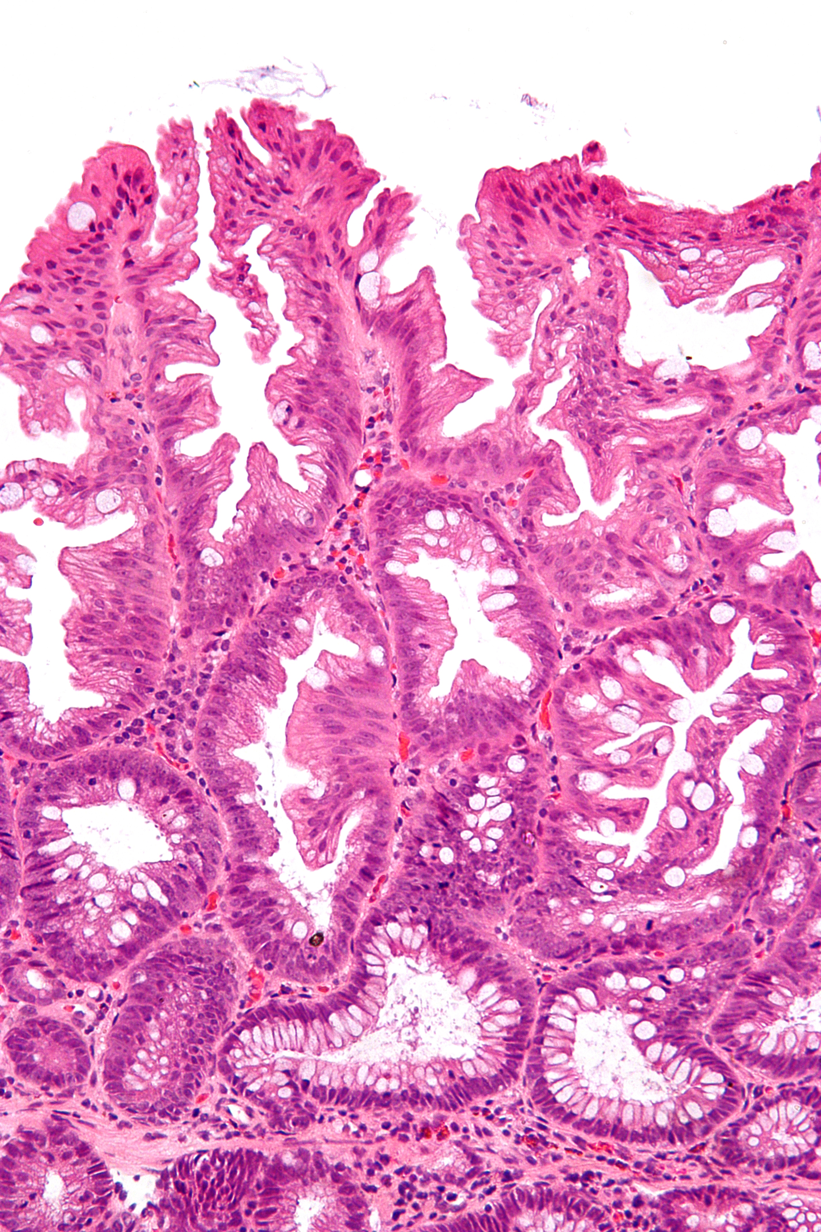
| Description | Very high magnification micrograph of a sessile serrated adenoma, abbreviated SSA, from the cecum removed during a colonoscopy. H&E stain.
SSAs are characterized by: basal dilation of the crypts, basal crypt serration, crypts that run horizontal to the basement membrane (horizontal crypts), and crypt branching. Unlike traditional colonic adenomas (e.g. tubular adenoma, villous adenoma), they do not typically have nuclear changes (nuclear hyperchromatism, nuclear crowding, elliptical/cigar-shaped nuclei). SSAs are considered pre-malignant lesions, i.e. precursors to cancer, and tend to be found in the right colon. Related images
Low mag.
Intermed. mag.
High mag.
Very high mag.
Low mag.
Intermed. mag.
|
| Source | Wikimedia Commons file page |
| Author | Nephron |
| Permission | See original Commons license details. |
Licensing[edit]
Creative Commons Attribution-ShareAlike 3.0 Unported (CC BY-SA 3.0)
This file is licensed under the Creative Commons Attribution-ShareAlike 3.0 license.
Official license: CC BY-SA 3.0
License page: CC BY-SA 3.0
Original attribution and file history: Wikimedia Commons
File history
Click on a date/time to view the file as it appeared at that time.
| Date/Time | Thumbnail | Dimensions | User | Comment | |
|---|---|---|---|---|---|
| current | 03:35, 5 June 2026 | | 2,848 × 4,272 (5.13 MB) | Maintenance script (talk | contribs) | == Summary == Importing file |
You cannot overwrite this file.
File usage
The following file is a duplicate of this file (more details):
- File:Sessile serrated adenoma 3 very high mag.jpg from Wikimedia Commons
The following 3 pages use this file:
