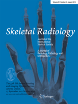
Skeletal Radiology
Skeletal Radiology is a specialized branch of radiology focusing on the diagnostic imaging of bones, joints, and associated soft tissues. It employs various imaging techniques to diagnose, treat, and manage conditions and diseases affecting the skeletal system. This field plays a crucial role in identifying fractures, infections, tumors, congenital anomalies, and degenerative diseases such as arthritis.
Overview[edit | edit source]
Skeletal Radiology utilizes a range of imaging modalities to visualize the anatomy and pathology of the skeletal system. The most commonly used techniques include:
- X-ray: The fundamental tool in skeletal radiology, providing high-resolution images of bone structure to detect fractures, infections, and tumors.
- Computed Tomography (CT): Offers detailed cross-sectional images, allowing for the assessment of complex fractures and bone diseases.
- Magnetic Resonance Imaging (MRI): Especially useful for imaging soft tissues related to the skeletal system, such as cartilage, muscles, and ligaments. MRI is invaluable in diagnosing soft tissue injuries, joint diseases, and bone marrow pathology.
- Ultrasound: Used for assessing soft tissue structures around the joints and can help in guiding joint injections or aspirations.
- Nuclear Medicine: Including bone scans, which can detect bone growth or repair, indicating fractures, cancer, or infections not yet visible on other imaging modalities.
Applications[edit | edit source]
Skeletal Radiology has a wide range of applications, including but not limited to:
- Diagnosis and management of fractures and dislocations
- Detection and monitoring of osteoporosis
- Evaluation of bone tumors and metastases
- Identification of inflammatory and degenerative joint diseases such as rheumatoid arthritis and osteoarthritis
- Guidance for biopsy or intervention procedures
- Preoperative planning and postoperative assessment
Challenges and Developments[edit | edit source]
The field of skeletal radiology is continuously evolving, with advancements in imaging technology and techniques enhancing diagnostic accuracy and patient care. However, challenges remain, such as the need for improved imaging resolution, faster scanning times, and reduced radiation exposure. Recent developments include the use of weight-bearing CT for foot and ankle imaging, high-resolution MRI for detailed soft tissue evaluation, and the application of artificial intelligence to assist in image analysis and diagnosis.
Education and Training[edit | edit source]
Becoming a skeletal radiologist requires extensive education and training. After completing medical school, a physician must undergo residency training in radiology, followed by a fellowship in musculoskeletal radiology. This specialized training covers all aspects of musculoskeletal imaging, including advanced MRI techniques, interventional procedures, and the use of ultrasound in musculoskeletal diagnosis.
Conclusion[edit | edit source]
Skeletal Radiology is an essential field within radiology, providing critical insights into the health and condition of the skeletal system. Through the use of advanced imaging techniques, skeletal radiologists play a vital role in diagnosing and managing a wide range of conditions, contributing significantly to patient care and treatment outcomes.
Navigation: Wellness - Encyclopedia - Health topics - Disease Index - Drugs - World Directory - Gray's Anatomy - Keto diet - Recipes
Search WikiMD
Ad.Tired of being Overweight? Try W8MD's physician weight loss program.
Semaglutide (Ozempic / Wegovy and Tirzepatide (Mounjaro / Zepbound) available.
Advertise on WikiMD
WikiMD is not a substitute for professional medical advice. See full disclaimer.
Credits:Most images are courtesy of Wikimedia commons, and templates Wikipedia, licensed under CC BY SA or similar.Contributors: Prab R. Tumpati, MD

