File:1gwe antipar betaSheet both.png
From WikiMD's WELLNESSPEDIA
Size of this preview: 800 × 440 pixels. Other resolution: 2,000 × 1,100 pixels.
Original file (2,000 × 1,100 pixels, file size: 1.04 MB, MIME type: image/png)
Summary[edit]
| Summary | |
|---|---|
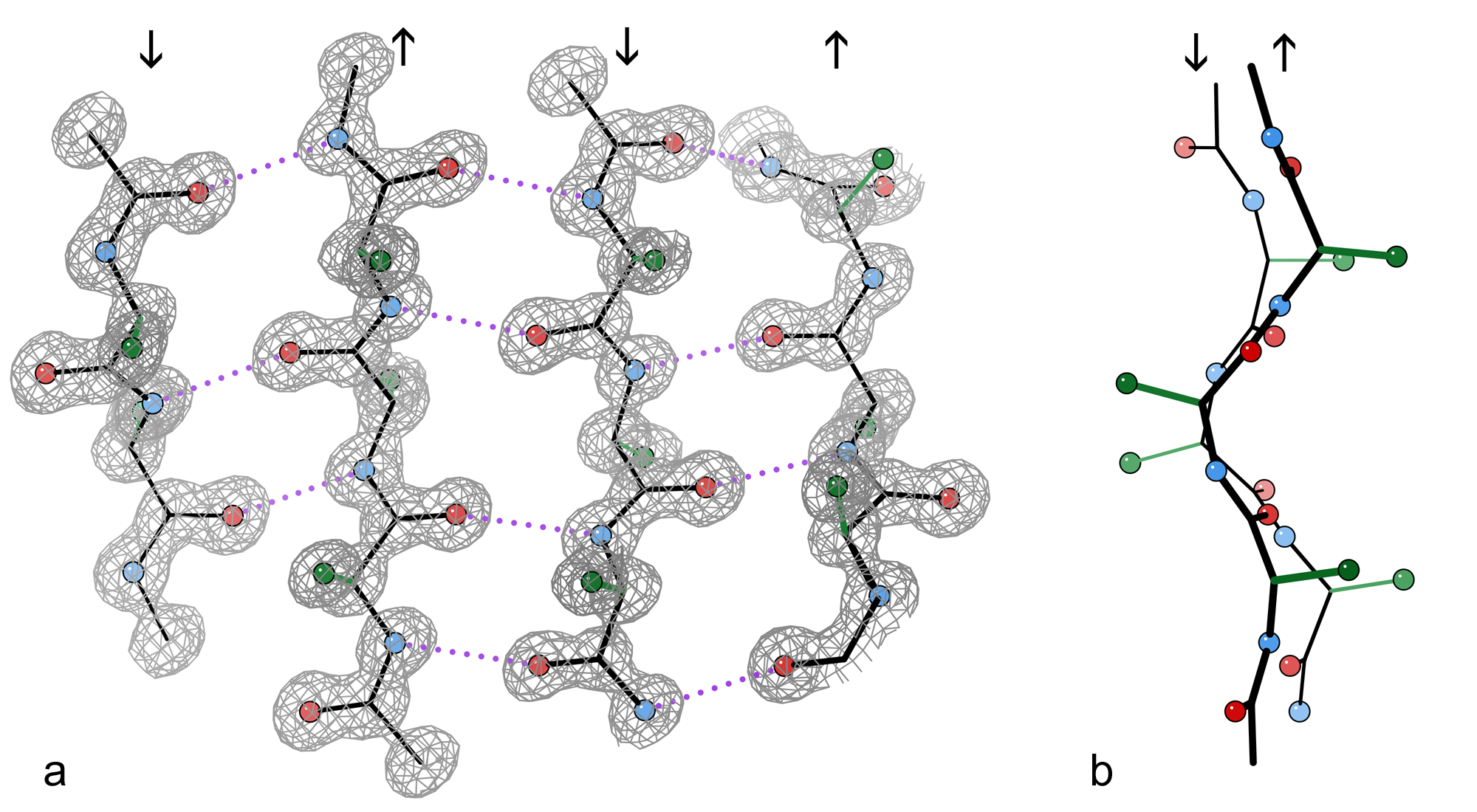
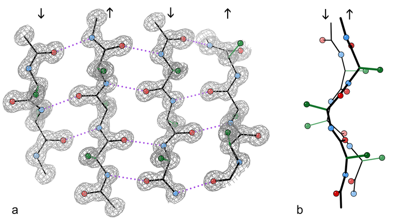
| Description | An example of a 4-stranded antiparallel β sheet fragment from a crystal structure of the enzyme catalase (PDB file 1GWE at 0.88 Å resolution). a) Front view, showing the antiparallel hydrogen bonds (dotted) between peptide NH and CO groups on adjacent strands. Arrows indicate chain direction, and electron density contours outline the non-H atoms. O atoms are red balls, N atoms are blue, and H atoms are omitted for simplicity; sidechains are shown only out to the first sidechain C atom (green). b) Edge-on view of the central two β strands in a, showing the righthanded twist and the pleat of Cαs and sidechains that alternately stick out in opposite directions from the sheet. |
| Source | Wikimedia Commons file page |
| Author | Dcrjsr |
| Permission | See original Commons license details. |
Licensing[edit]
Creative Commons Attribution 3.0 Unported (CC BY 3.0)
This file is licensed under the Creative Commons Attribution 3.0 license.
Official license: CC BY 3.0
License page: CC BY 3.0
Original attribution and file history: Wikimedia Commons
File history
Click on a date/time to view the file as it appeared at that time.
| Date/Time | Thumbnail | Dimensions | User | Comment | |
|---|---|---|---|---|---|
| current | 02:08, 7 June 2026 | | 2,000 × 1,100 (1.04 MB) | Maintenance script (talk | contribs) | == Summary == Importing file |
You cannot overwrite this file.
File usage
The following file is a duplicate of this file (more details):
- File:1gwe antipar betaSheet both.png from Wikimedia Commons
The following page uses this file:
