File:3D-SIM-1 NPC Confocal vs 3D-SIM.jpg
From WikiMD's WELLNESSPEDIA
Size of this preview: 756 × 600 pixels. Other resolution: 1,069 × 848 pixels.
Original file (1,069 × 848 pixels, file size: 307 KB, MIME type: image/jpeg)
Summary[edit]
| Summary | |
|---|---|
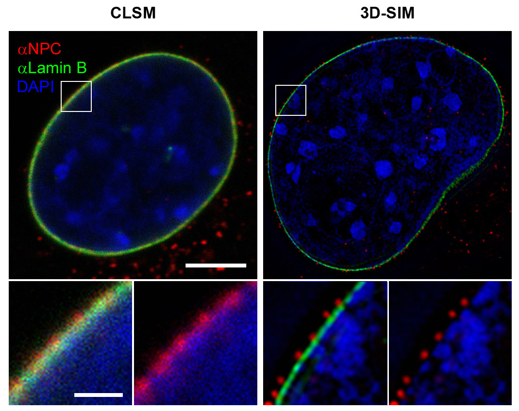
| Description | Comparison of resolution obtained by confocal laser scanning microscopy (clsm, left) and 3D structured illumination microscopy (3D-SIM-Microscopy, right). Top images each show a nucleus of a mouse cell, the white boxes are magnified at the bottom. Nuclear pores (anti-NPC, red) nuclear envelope (anti-Lamin, green). Chromatin/DNA (DAPI, blue). scale bars: top 5 µm, bottom 1µm. For further information see: Schermelleh L, Carlton PM, Haase S, Shao L, Winoto L, Kner P, Burke B, Cardoso MC, Agard DA, Gustafsson MG, Leonhardt H, Sedat JW (June 2008). "Subdiffraction multicolor imaging of the nuclear periphery with 3D structured illumination microscopy". Science (journal) 320 (5881): 1332–6. DOI:10.1126/science.1156947. PMID 18535242. |
| Source | Wikimedia Commons file page |
| Author | Lothar Schermelleh |
| Permission | See original Commons license details. |
Licensing[edit]
Creative Commons Attribution-ShareAlike 3.0 Unported (CC BY-SA 3.0)
This file is licensed under the Creative Commons Attribution-ShareAlike 3.0 license.
Official license: CC BY-SA 3.0
License page: CC BY-SA 3.0
Original attribution and file history: Wikimedia Commons
File history
Click on a date/time to view the file as it appeared at that time.
| Date/Time | Thumbnail | Dimensions | User | Comment | |
|---|---|---|---|---|---|
| current | 02:12, 7 June 2026 | | 1,069 × 848 (307 KB) | Maintenance script (talk | contribs) | == Summary == Importing file |
You cannot overwrite this file.
File usage
The following page uses this file:
