File:3D-SIM-2 Nucleus prophase 3d rotated.jpg
From WikiMD's WELLNESSPEDIA
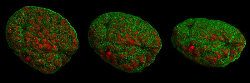
Size of this preview: 800 × 267 pixels. Other resolution: 1,920 × 640 pixels.
Original file (1,920 × 640 pixels, file size: 377 KB, MIME type: image/jpeg)
Summary[edit]
| Summary | |
|---|---|
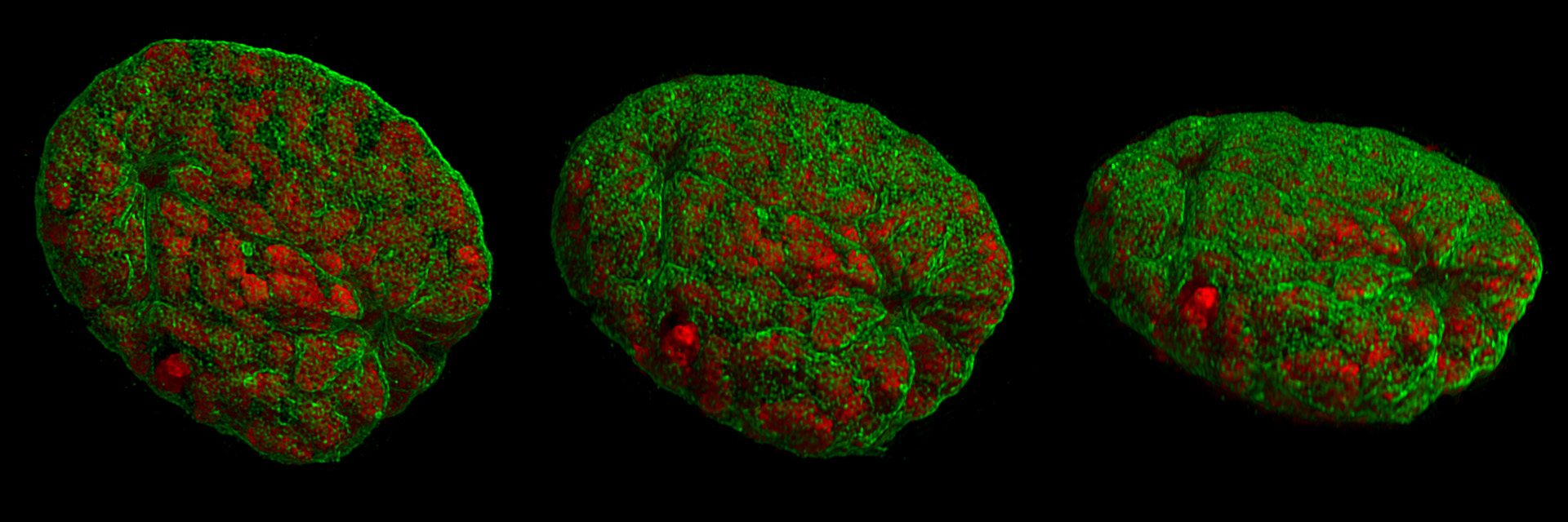
| Description | 3D-representation of a mouse cell nucleus from different angles. Recorded with 3D Structured Illumination Microscopy (3D-SIM-microscopy). The cell is in an early stage of nuclear division (prophase). The chromosomes (red) are already condensed, to be distributed to the daughter cells later on. The surrounding nuclear envelope (green) shows prominent invaginations and first disruptions. For further information see: Schermelleh L, Carlton PM, Haase S, Shao L, Winoto L, Kner P, Burke B, Cardoso MC, Agard DA, Gustafsson MG, Leonhardt H, Sedat JW (June 2008). "Subdiffraction multicolor imaging of the nuclear periphery with 3D structured illumination microscopy". Science (journal) 320 (5881): 1332–6. DOI:10.1126/science.1156947. PMID 18535242. |
| Source | Wikimedia Commons file page |
| Author | Lothar Schermelleh |
| Permission | See original Commons license details. |
Licensing[edit]
Creative Commons Attribution-ShareAlike 3.0 Unported (CC BY-SA 3.0)
This file is licensed under the Creative Commons Attribution-ShareAlike 3.0 license.
Official license: CC BY-SA 3.0
License page: CC BY-SA 3.0
Original attribution and file history: Wikimedia Commons
File history
Click on a date/time to view the file as it appeared at that time.
| Date/Time | Thumbnail | Dimensions | User | Comment | |
|---|---|---|---|---|---|
| current | 02:07, 7 June 2026 | 1,920 × 640 (377 KB) | Maintenance script (talk | contribs) | == Summary == Importing file |
You cannot overwrite this file.
File usage
The following page uses this file:
