File:3D-SIM-3 Prophase 3 color.jpg
From WikiMD's WELLNESSPEDIA
Size of this preview: 604 × 600 pixels. Other resolution: 902 × 896 pixels.
Original file (902 × 896 pixels, file size: 386 KB, MIME type: image/jpeg)
Summary[edit]
| Summary | |
|---|---|
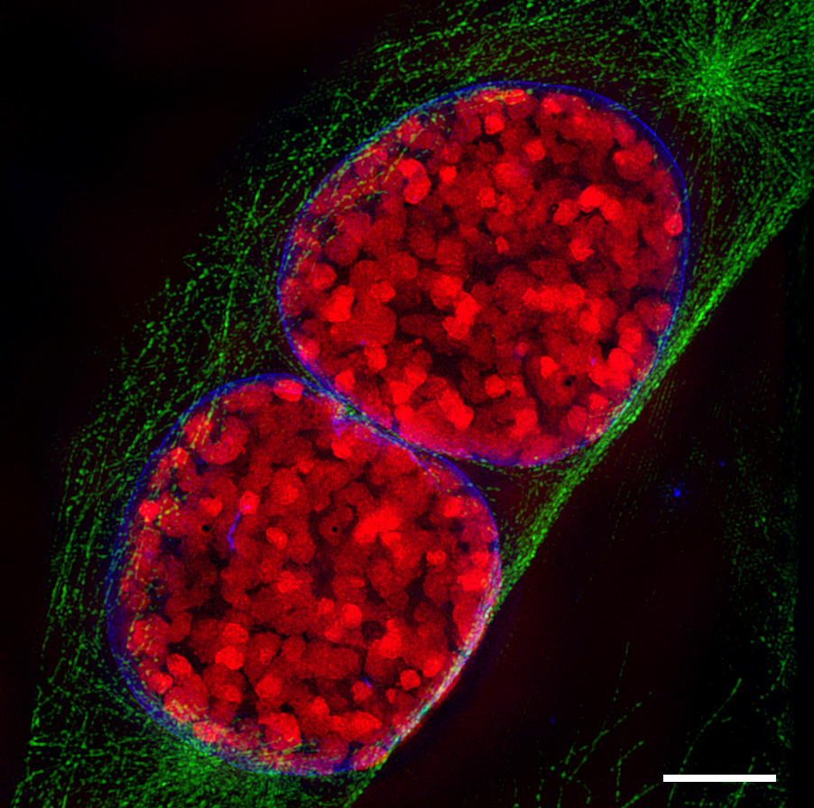
| Description | Light-optical section through two mouse cell nuclei in prophase, recorded with 3D Structured Illumination Microscopy (3D-SIM-microscopy). condensed chromosomes are red, the nuclear envelope blue and microtubuli, which belong to the cytoskeleton, are green. Scale bar is 5 µm. For further information see: Schermelleh L, Carlton PM, Haase S, Shao L, Winoto L, Kner P, Burke B, Cardoso MC, Agard DA, Gustafsson MG, Leonhardt H, Sedat JW (June 2008). "Subdiffraction multicolor imaging of the nuclear periphery with 3D structured illumination microscopy". Science (journal) 320 (5881): 1332–6. DOI:10.1126/science.1156947. PMID 18535242. |
| Source | Wikimedia Commons file page |
| Author | Lothar Schermelleh |
| Permission | See original Commons license details. |
Licensing[edit]
Creative Commons Attribution-ShareAlike 3.0 Unported (CC BY-SA 3.0)
This file is licensed under the Creative Commons Attribution-ShareAlike 3.0 license.
Official license: CC BY-SA 3.0
Original attribution and file history: Wikimedia Commons
File history
Click on a date/time to view the file as it appeared at that time.
| Date/Time | Thumbnail | Dimensions | User | Comment | |
|---|---|---|---|---|---|
| current | 12:52, 29 May 2026 | | 902 × 896 (386 KB) | Maintenance script (talk | contribs) | == Summary == Importing file |
You cannot overwrite this file.
File usage
The following file is a duplicate of this file (more details):
- File:3D-SIM-3 Prophase 3 color.jpg from Wikimedia Commons
The following 2 pages use this file:
