File:405663-PLEOMORPHIC XANTHOASTROCYTOMA.jpg
From WikiMD's WELLNESSPEDIA
No higher resolution available.
405663-PLEOMORPHIC_XANTHOASTROCYTOMA.jpg (421 × 512 pixels, file size: 125 KB, MIME type: image/jpeg)
Summary[edit]
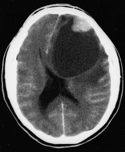
| Summary | |
|---|---|
| Description | PLEOMORPHIC XANTHOASTROCYTOMA As in this computerized tomographic scan, the classic radiographic appearance is one of a superficially situated tumor, here a mural nodule, associated with an underlying cyst. |
| Source | Wikimedia Commons file page |
| Author | The Armed Forces Institute of Pathology |
| Permission | See original Commons license details. |
Licensing[edit]
Public Domain
This file is in the public domain and may be used without restriction.
Please see the linked source page for the original file history, attribution information, and licensing details.
Original attribution and file history: Wikimedia Commons
File history
Click on a date/time to view the file as it appeared at that time.
| Date/Time | Thumbnail | Dimensions | User | Comment | |
|---|---|---|---|---|---|
| current | 02:08, 7 June 2026 | | 421 × 512 (125 KB) | Maintenance script (talk | contribs) | == Summary == Importing file |
You cannot overwrite this file.
File usage
The following file is a duplicate of this file (more details):
- File:405663-PLEOMORPHIC XANTHOASTROCYTOMA.jpg from Wikimedia Commons
The following page uses this file:
