File:41467 2020 20149 Fig1f.jpg
From WikiMD's WELLNESSPEDIA
Size of this preview: 472 × 599 pixels. Other resolution: 781 × 991 pixels.
Original file (781 × 991 pixels, file size: 311 KB, MIME type: image/jpeg)
Summary[edit]
| Summary | |
|---|---|
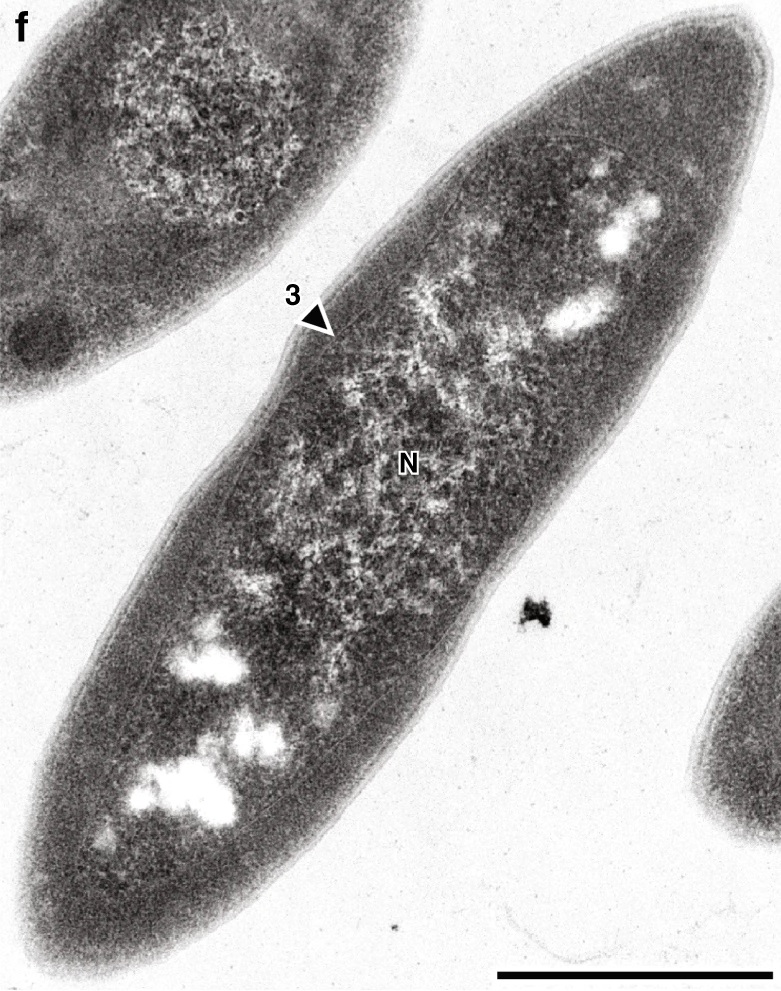
| Description | Morphology and membrane structure in Atribacter laminatus (type strain RT761) cells showing the presence of three lipid membrane-like layers (LMLs) with the innermost layer surrounding the nucleoid. Transmission electron micrograph of a thin section of RT761 cells. N nucleoid. Scale bar, 0.5 μm. |
| Source | Wikimedia Commons file page |
| Author | Taiki Katayama, Masaru K. Nobu, Hiroyuki Kusada, Xian-Ying Meng, Naoki Hosogi, Katsuyuki Uematsu, Hideyoshi Yoshioka, Yoichi Kamagata, Hideyuki Tamaki |
| Permission | See original Commons license details. |
Licensing[edit]
Creative Commons Attribution-ShareAlike 4.0 International (CC BY-SA 4.0)
This file is licensed under the Creative Commons Attribution-ShareAlike 4.0 International license.
You are free to:
- Share — copy and redistribute the material.
- Adapt — remix, transform, and build upon the material.
Under the following conditions:
- Attribution — appropriate credit must be given.
- ShareAlike — derivative works must be distributed under the same license.
Official license: CC BY-SA 4.0
License page: CC BY-SA 4.0
Original attribution and file history: Wikimedia Commons
File history
Click on a date/time to view the file as it appeared at that time.
| Date/Time | Thumbnail | Dimensions | User | Comment | |
|---|---|---|---|---|---|
| current | 02:09, 7 June 2026 | | 781 × 991 (311 KB) | Maintenance script (talk | contribs) | == Summary == Importing file |
You cannot overwrite this file.
File usage
The following page uses this file:
