File:4OOG.png
Original file (1,800 × 900 pixels, file size: 1.1 MB, MIME type: image/png)
Summary[edit]
| Summary | |
|---|---|
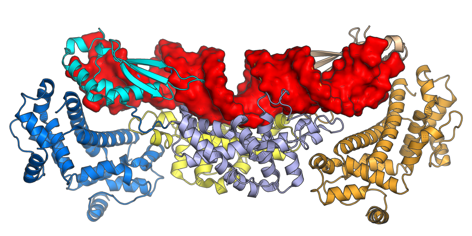
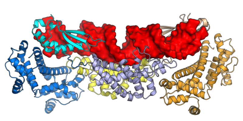
| Description | The crystal structure of the class I ribonuclease III (Rnt1p) from Saccharomyces cerevisiae in complex with double-stranded RNA (shown in surface representation in red). The protein is a dimer; the N-terminal domains are shown in blue and orange; the dsRNA-binding domains in cyan and tan; and the ribonuclease domains in light blue and yellow.
Rendered from PDB 4OOG. Liang YH, Lavoie M, Comeau MA, Elela SA, Ji X. Structure of a eukaryotic RNase III postcleavage complex reveals a double-ruler mechanism for substrate selection. Molecular cell. 2014 May 8;54(3):431-44. DOI: 10.1016/j.molcel.2014.03.006 |
| Source | Wikimedia Commons file page |
| Author | Opabinia regalis |
| Permission | See original Commons license details. |
Licensing[edit]
Creative Commons Attribution-ShareAlike 4.0 International (CC BY-SA 4.0)
This file is licensed under the Creative Commons Attribution-ShareAlike 4.0 International license.
You are free to:
- Share — copy and redistribute the material.
- Adapt — remix, transform, and build upon the material.
Under the following conditions:
- Attribution — appropriate credit must be given.
- ShareAlike — derivative works must be distributed under the same license.
Official license: CC BY-SA 4.0
License page: CC BY-SA 4.0
Original attribution and file history: Wikimedia Commons
File history
Click on a date/time to view the file as it appeared at that time.
| Date/Time | Thumbnail | Dimensions | User | Comment | |
|---|---|---|---|---|---|
| current | 02:08, 7 June 2026 | | 1,800 × 900 (1.1 MB) | Maintenance script (talk | contribs) | == Summary == Importing file |
You cannot overwrite this file.
File usage
The following page uses this file:
