File:6VSB spike protein SARS-CoV-2 monomer in homotrimer.png
From WikiMD's WELLNESSPEDIA
Size of this preview: 397 × 599 pixels. Other resolution: 989 × 1,491 pixels.
Original file (989 × 1,491 pixels, file size: 241 KB, MIME type: image/png)
Summary[edit]
| Summary | |
|---|---|
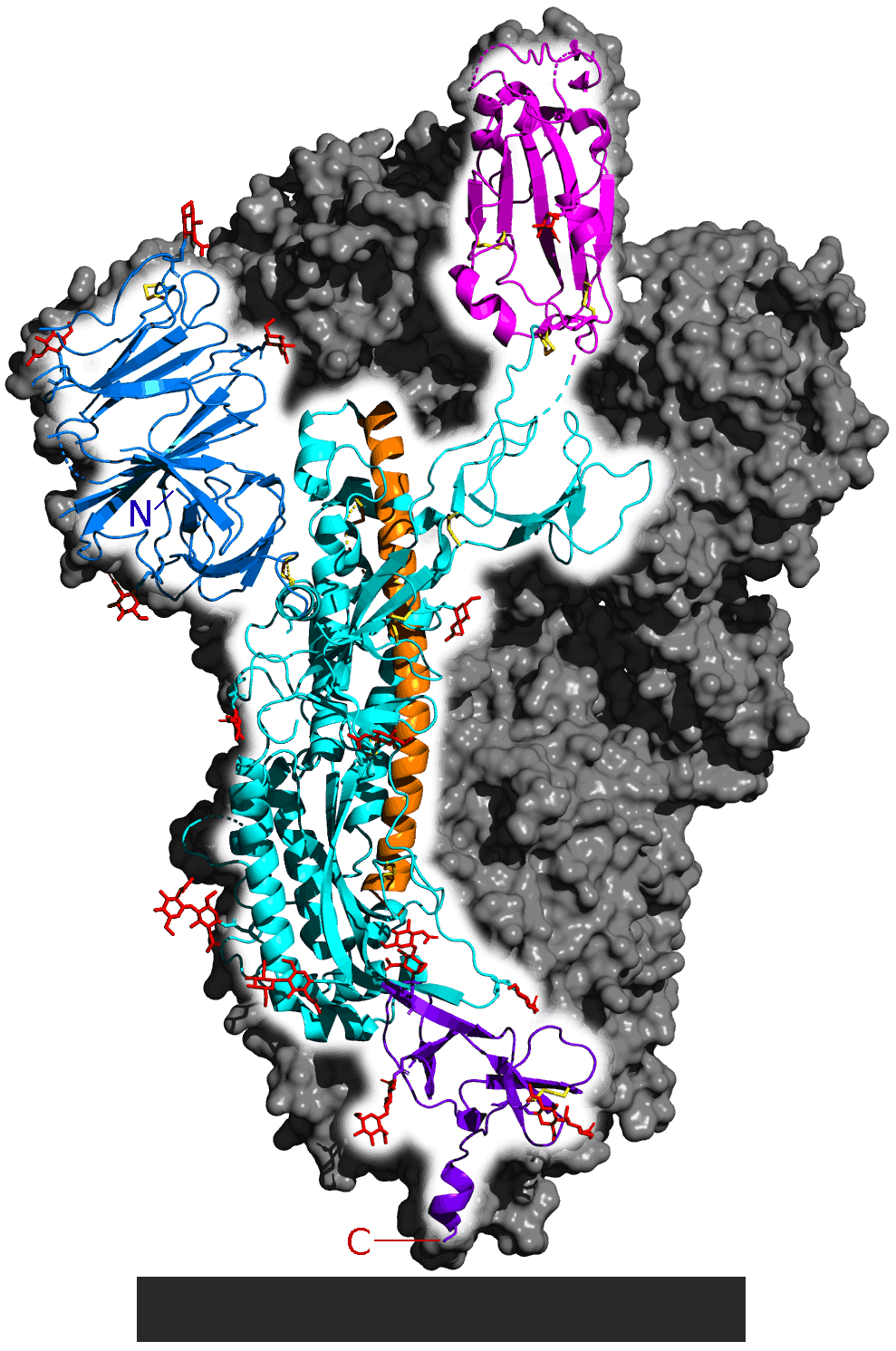
| Description | Spike glycoprotein from SARS-CoV-2. PDB: 6VSB. Only one monomer is highlighted. Whole protein is a homotrimer. Rest of the trimer is shown as a gray surface. Parts of the actual structure are not shown. The following are listed from N-terminal (letter N) to C-terminal (C): N-terminal domain (blue), ACE2 receptor binding domain (magenta) general structure (cyan), central helix (orange, faces inside of the homotrimer) and connector domain (purple, anchors the spike protein to virus lipid envelope). Yellow: disulfide bonds. Red: carbohydrates. Gray block: lipid membrane of the virus. |
| Source | Wikimedia Commons file page |
| Author | 5-HT2AR |
| Permission | See original Commons license details. |
Licensing[edit]
License: CC0
License page: CC0
Original attribution and file history: Wikimedia Commons
File history
Click on a date/time to view the file as it appeared at that time.
| Date/Time | Thumbnail | Dimensions | User | Comment | |
|---|---|---|---|---|---|
| current | 02:11, 7 June 2026 | | 989 × 1,491 (241 KB) | Maintenance script (talk | contribs) | == Summary == Importing file |
You cannot overwrite this file.
File usage
The following file is a duplicate of this file (more details):
- File:6VSB spike protein SARS-CoV-2 monomer in homotrimer.png from Wikimedia Commons
The following page uses this file:
