File:A computed tomography brain scan showing bilateral basal ganglia calcification.jpg
From WikiMD's WELLNESSPEDIA
Size of this preview: 800 × 589 pixels. Other resolution: 1,200 × 883 pixels.
Original file (1,200 × 883 pixels, file size: 153 KB, MIME type: image/jpeg)
Summary[edit]
| Summary | |
|---|---|
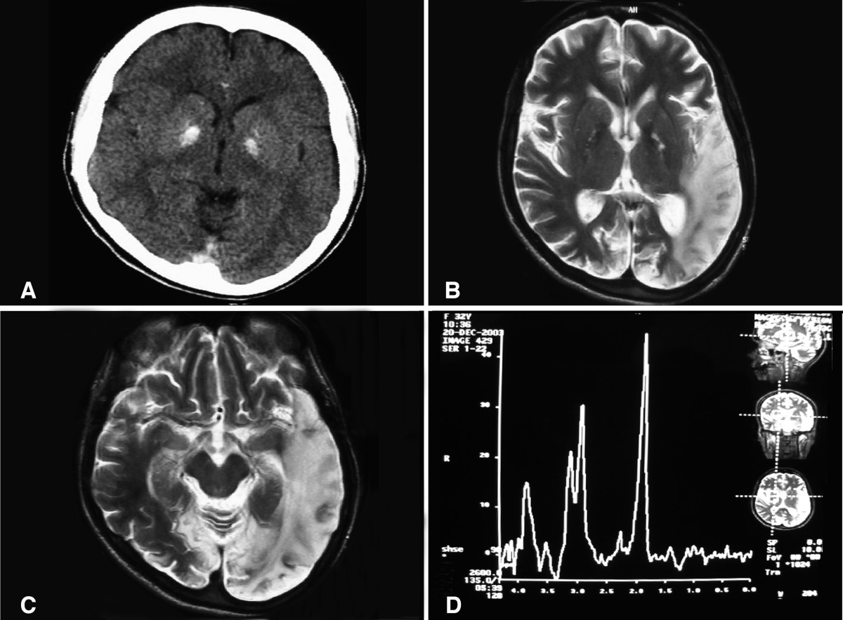
| Description | (a) A computed tomography brain scan showing bilateral basal ganglia calcification; the cerebellum shows prominent folia indicating mild cerebellar atrophy. (b) Axial T2 brain magnetic resonance image scan showing left temporo-parieto occipital ischemic lesion. (c) Axial T2 brain magnetic resonance image scan showing the extension of the parietal temporal region to the occipital lobe, and also showing a right occipital lesion. (d) Magnetic resonance spectroscopy showing inversion of J-coupling phenomenon at 1.3 ppm, indicating lactate peak. Abu-Amero et al. Journal of Medical Case Reports 2009 3:77 doi:10.1186/1752-1947-3-77 |
| Source | Wikimedia Commons file page |
| Author | Abu-Amero KK, Al-Dhalaan H, Bohlega S, Hellani A, Taylor RW. |
| Permission | See original Commons license details. |
Licensing[edit]
License: CC BY 2.0
License page: CC BY 2.0
Original attribution and file history: Wikimedia Commons
File history
Click on a date/time to view the file as it appeared at that time.
| Date/Time | Thumbnail | Dimensions | User | Comment | |
|---|---|---|---|---|---|
| current | 02:09, 7 June 2026 | | 1,200 × 883 (153 KB) | Maintenance script (talk | contribs) | == Summary == Importing file |
You cannot overwrite this file.
File usage
The following file is a duplicate of this file (more details):
- File:A computed tomography brain scan showing bilateral basal ganglia calcification.jpg from Wikimedia Commons
The following 3 pages use this file:
