File:Assembly of fibrous muscle, fat, and vascular tissues to cultured steak.webp
From WikiMD's WELLNESSPEDIA
Size of this PNG preview of this WEBP file: 800 × 482 pixels.
Original file (1,499 × 904 pixels, file size: 182 KB, MIME type: image/webp)
Summary[edit]
| Summary | |
|---|---|
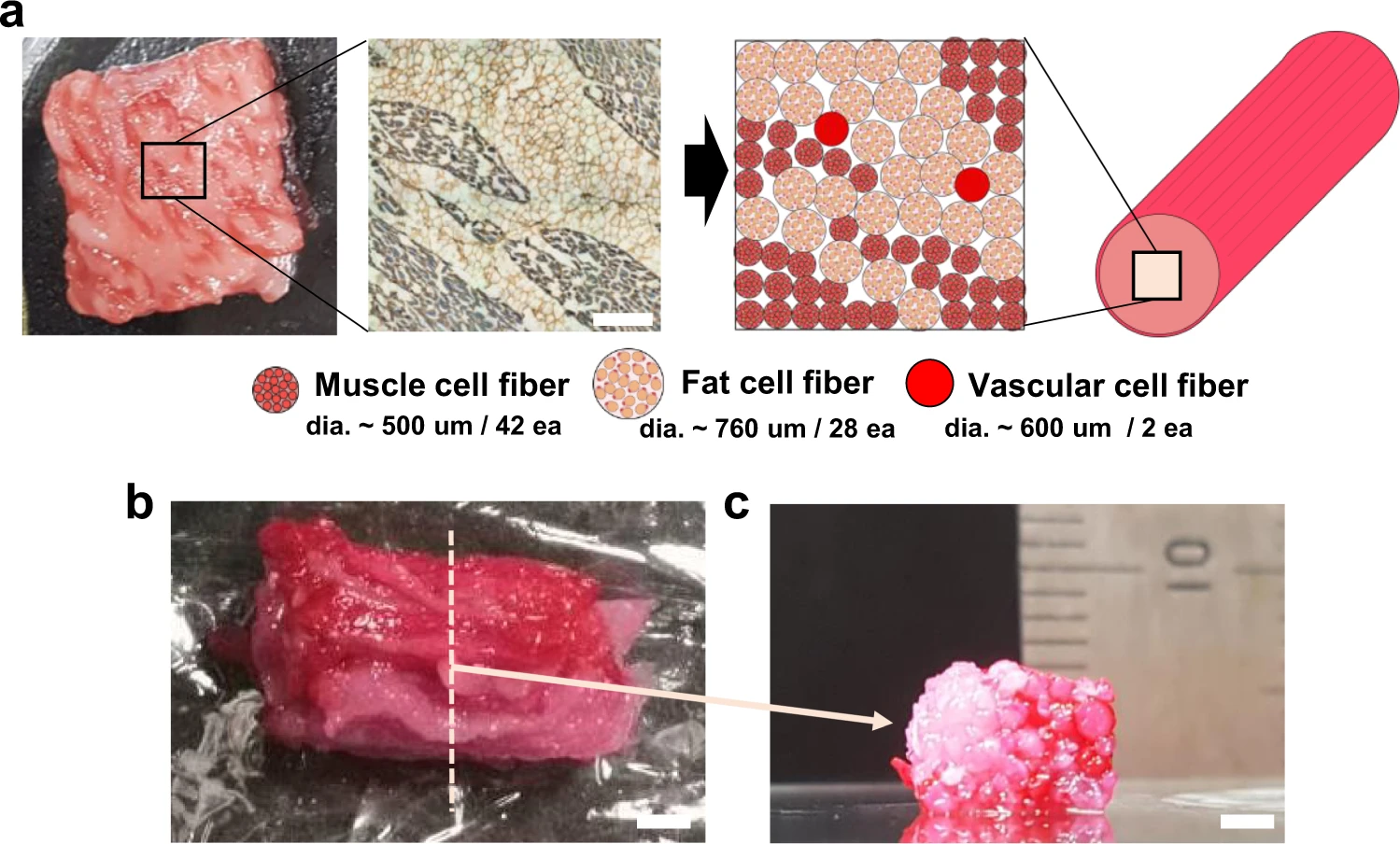
| Description | "a Assembly schematic- (right) based sarcomeric α-actinin (blue) and laminin- (brown) stained image (left) of the commercial meat. It is assumed that the diameters of the fibrous muscle, fat, and vascular tissues are about 500, 760, and 600 µm, respectively. Scale bar, 1 mm. b, c Optical images of the cultured steak by assembling muscle (42 ea.), fat (28 ea.), and vascular (2 ea.) tissues at (b) the top and (c) cross-section view of the dotted-line area. Muscle and vascular tissue were stained with carmine (red color), but fat tissue was not. Scale bars, 2 mm." |
| Source | Wikimedia Commons file page |
| Author | Authors of the study: Dong-Hee Kang, Fiona Louis, Hao Liu, Hiroshi Shimoda, Yasutaka Nishiyama, Hajime Nozawa, Makoto Kakitani, Daisuke Takagi, Daijiro Kasa, Eiji Nagamori, Shinji Irie, Shiro Kitano & Michiya Matsusaki |
| Permission | See original Commons license details. |
Licensing[edit]
Creative Commons Attribution 4.0 International (CC BY 4.0)
This file is licensed under the Creative Commons Attribution 4.0 International license.
You may share and adapt the material provided appropriate attribution is given.
Official license: CC BY 4.0
License page: CC BY 4.0
Original attribution and file history: Wikimedia Commons
File history
Click on a date/time to view the file as it appeared at that time.
| Date/Time | Thumbnail | Dimensions | User | Comment | |
|---|---|---|---|---|---|
| current | 03:21, 5 June 2026 | | 1,499 × 904 (182 KB) | Maintenance script (talk | contribs) | == Summary == Importing file |
You cannot overwrite this file.
File usage
The following file is a duplicate of this file (more details):
- File:Assembly of fibrous muscle, fat, and vascular tissues to cultured steak.webp from Wikimedia Commons
The following 2 pages use this file:
