File:Bilateral Sciatic Neurography.jpg
From WikiMD's WELLNESSPEDIA
No higher resolution available.
Bilateral_Sciatic_Neurography.jpg (586 × 557 pixels, file size: 212 KB, MIME type: image/jpeg)
Summary[edit]
| Summary | |
|---|---|
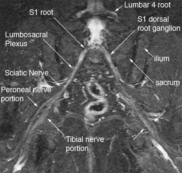
| Description | Magnetic Resonance Neurography image of the posterior pelvis showing the S1 nerve roots, the dorsal root ganglia, the lumbosacral plexus, and the proximal sciatic nerve forming at the sciatic notch. This individual has an anatomical variant in which the peroneal and tibial portions of the sciatic nerve are split as they pass through the piriformis muscle. The image was obtained on a GE 1.5 Tesla MRI scanner with phased array imaging coils. |
| Source | Wikimedia Commons file page |
| Author | Aaron FillerAfiller (talk) |
| Permission | See original Commons license details. |
Licensing[edit]
Creative Commons Attribution-ShareAlike 3.0 Unported (CC BY-SA 3.0)
This file is licensed under the Creative Commons Attribution-ShareAlike 3.0 license.
Official license: CC BY-SA 3.0
License page: CC BY-SA 3.0
Original attribution and file history: Wikimedia Commons
File history
Click on a date/time to view the file as it appeared at that time.
| Date/Time | Thumbnail | Dimensions | User | Comment | |
|---|---|---|---|---|---|
| current | 03:18, 5 June 2026 | | 586 × 557 (212 KB) | Maintenance script (talk | contribs) | == Summary == Importing file |
You cannot overwrite this file.
File usage
The following 2 pages use this file:
