File:Brodmann areas 6.png
Brodmann_areas_6.png (256 × 192 pixels, file size: 28 KB, MIME type: image/png)
Summary[edit]
| Summary | |
|---|---|
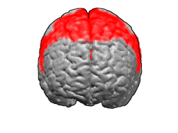
| Description | Brodmann areas 6
BA6 is in the posterior part of the frontal lobe. This is a front view, looking down on the brain The brain's surface is extracted from structural MRI data (Wellcome Dept. Imaging Neuroscience, UCL, UK). The Brodmann Area data is based on information from the online Talairach demon (an electronic version of Talairach and Tournoux, 1988). These images were created using Blender and Matlab. reference:Talairach, J., and Tournoux, P. Co-Planar Stereotactic Atlas of the Human Brain., New York: Thieme, 1988. |
| Source | Wikimedia Commons file page |
| Author | Washington irving |
| Permission | See original Commons license details. |
Licensing[edit]
Creative Commons Attribution-ShareAlike 3.0 Unported (CC BY-SA 3.0)
This file is licensed under the Creative Commons Attribution-ShareAlike 3.0 license.
Official license: CC BY-SA 3.0
License page: CC BY-SA 3.0
Original attribution and file history: Wikimedia Commons
File history
Click on a date/time to view the file as it appeared at that time.
| Date/Time | Thumbnail | Dimensions | User | Comment | |
|---|---|---|---|---|---|
| current | 03:33, 5 June 2026 | | 256 × 192 (28 KB) | Maintenance script (talk | contribs) | == Summary == Importing file |
You cannot overwrite this file.
File usage
The following file is a duplicate of this file (more details):
- File:Brodmann areas 6.png from Wikimedia Commons
The following 2 pages use this file:
