File:CT of lymphocytic interstitial pneumonia.jpg
From WikiMD's WELLNESSPEDIA

Size of this preview: 800 × 328 pixels. Other resolution: 2,700 × 1,106 pixels.
Original file (2,700 × 1,106 pixels, file size: 267 KB, MIME type: image/jpeg)
Summary[edit]
| Summary | |
|---|---|
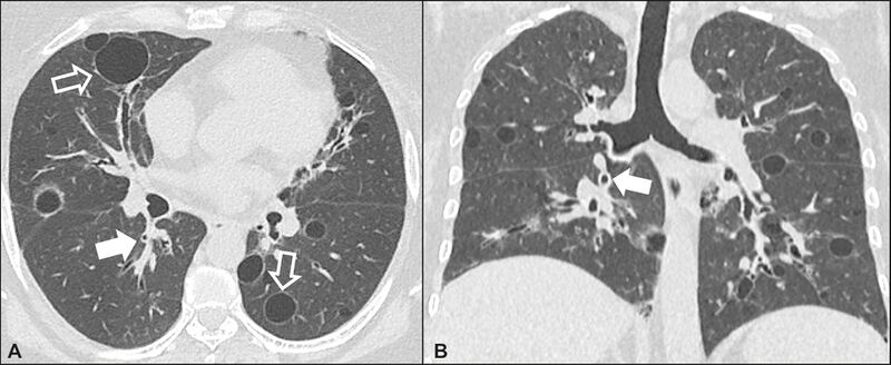
| Description | CT scan of lymphocytic interstitial pneumonia. Original caption: Figure 7 Lymphocytic interstitial pneumonia. A 62-year-old female patient with Sjögren’s syndrome. Axial high-resolution computed tomography scan of the chest (A) and coronal reformatting (B). In A, diffuse thickening of the bronchial walls (closed arrows), some ground-glass opacities and thin-walled cysts of varying sizes, with a diffuse, bilateral distribution (open arrows). In B, distribution predominantly in the lower fields. |
| Source | Wikimedia Commons file page |
| Author | Article authors: Daniel Simões Oliveira, José de Arimatéia Araújo Filho, Antonio Fernando Lins Paiva, Eduardo Seigo Ikari, Rodrigo Caruso Chate, César Higa Nomura |
| Permission | See original Commons license details. |
Licensing[edit]
Creative Commons Attribution 4.0 International (CC BY 4.0)
This file is licensed under the Creative Commons Attribution 4.0 International license.
You may share and adapt the material provided appropriate attribution is given.
Official license: CC BY 4.0
License page: CC BY 4.0
Original attribution and file history: Wikimedia Commons
File history
Click on a date/time to view the file as it appeared at that time.
| Date/Time | Thumbnail | Dimensions | User | Comment | |
|---|---|---|---|---|---|
| current | 03:35, 5 June 2026 | 2,700 × 1,106 (267 KB) | Maintenance script (talk | contribs) | == Summary == Importing file |
You cannot overwrite this file.
File usage
The following 2 pages use this file:
