File:Centrocyte, centroblast and follicular dendritic cell in a follicular lymphoma.jpg
From WikiMD's WELLNESSPEDIA
No higher resolution available.
Centrocyte,_centroblast_and_follicular_dendritic_cell_in_a_follicular_lymphoma.jpg (427 × 301 pixels, file size: 52 KB, MIME type: image/jpeg)
Summary[edit]
| Summary | |
|---|---|
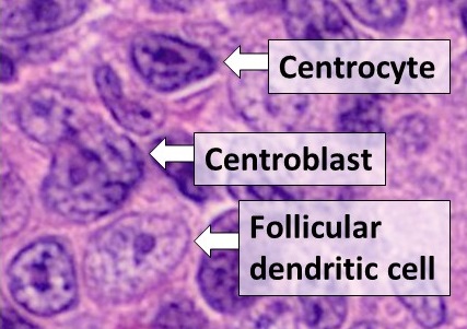
| Description | Histology of cell types in a germinal center: A centrocyte, a centroblast and a follicular dendritic cell, seen in a follicular lymphoma, H&E stain.- Centrocytes are small to medium size with angulated, elongated, cleaved, or twisted nuclei.- Centroblasts are larger cells containing vesicular nuclei with one to three basophilic nucleoli apposing the nuclear membrane.- Follicular dendritic cells have round nuclei, centrally located nucleoli, bland and dispersed chromatin, and flattening of adjacent nuclear membrane. Reference: Dr. William Kern. Case No.: H-003. Diagnosis: Non-Hodgkin lymphoma, follicular type, low-grade. Organ: Lymph node, inguinal. University of Oklahoma Health Sciences Center, Oklahoma. Last Updated: 12/21/2010 |
| Source | Wikimedia Commons file page |
| Author | Mikael Häggström, M.D. Author info - Reusing images- Conflicts of interest: None Mikael Häggström, M.D.Consent note: Consent from the patient or patient's relatives is regarded as redundant, because of absence of identifiable features (List of HIPAA identifiers) in the media and case information (See also HIPAA case reports guidance). |
| Permission | See original Commons license details. |
Licensing[edit]
License: CC0
License page: CC0
Original attribution and file history: Wikimedia Commons
File history
Click on a date/time to view the file as it appeared at that time.
| Date/Time | Thumbnail | Dimensions | User | Comment | |
|---|---|---|---|---|---|
| current | 03:25, 5 June 2026 | | 427 × 301 (52 KB) | Maintenance script (talk | contribs) | == Summary == Importing file |
You cannot overwrite this file.
File usage
The following file is a duplicate of this file (more details):
- File:Centrocyte, centroblast and follicular dendritic cell in a follicular lymphoma.jpg from Wikimedia Commons
The following 2 pages use this file:
