File:Cervical XRayFlexionExtension.jpg
Original file (2,800 × 2,008 pixels, file size: 355 KB, MIME type: image/jpeg)
Summary[edit]
| Summary | |
|---|---|
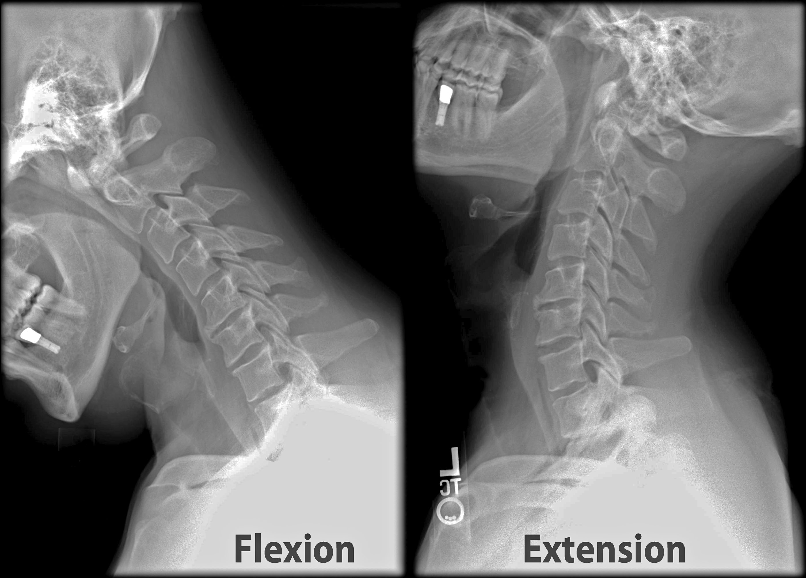
| Description | X-ray of cervical spine (neck) in flexion and extension (bending backwards). This series of x-rays were part of pre-surgical evaluation to help identify spinal instability. Patient is a 37 year old male with a history of multiple neck traumas with pain and muscle spasms and dental implant in lower jaw. Excerpt from radiologist's report:
FINDINGS: Five views of the cervical spine, including flexion and extension, were performed. There is no evidence of fracture, bone destruction, or malalignment. There are degenerative bone and is changes at C5-6. There is no evidence of cervical instability on the flexion and extension views. The facet joints are well aligned. Bony spurring is narrowing the C5-6 neural foramina bilaterally. IMPRESSION: Degenerative changes at C5-6. No evidence of instability. |
| Source | Wikimedia Commons |
| Author | Cervical_Xray_Extension.jpg: Stillwaterising
Cervical_Xray_Extension_view.jpg: Stillwaterising derivative work: F. Lamiot (talk) |
| Permission | See Commons |
Licensing[edit]
Creative Commons Attribution-ShareAlike 3.0 Unported (CC BY-SA 3.0)
This file is licensed under the Creative Commons Attribution-ShareAlike 3.0 license.
Official license: CC BY-SA 3.0
Original attribution and file history: Wikimedia Commons
File history
Click on a date/time to view the file as it appeared at that time.
| Date/Time | Thumbnail | Dimensions | User | Comment | |
|---|---|---|---|---|---|
| current | 01:20, 2 June 2026 | | 2,800 × 2,008 (355 KB) | Maintenance script (talk | contribs) | == Summary == Importing file |
You cannot overwrite this file.
File usage
The following 2 pages use this file:
