File:Chorioamnionitis - intermed mag.jpg
Original file (4,272 × 2,848 pixels, file size: 4.62 MB, MIME type: image/jpeg)
Summary[edit]
| Summary | |
|---|---|
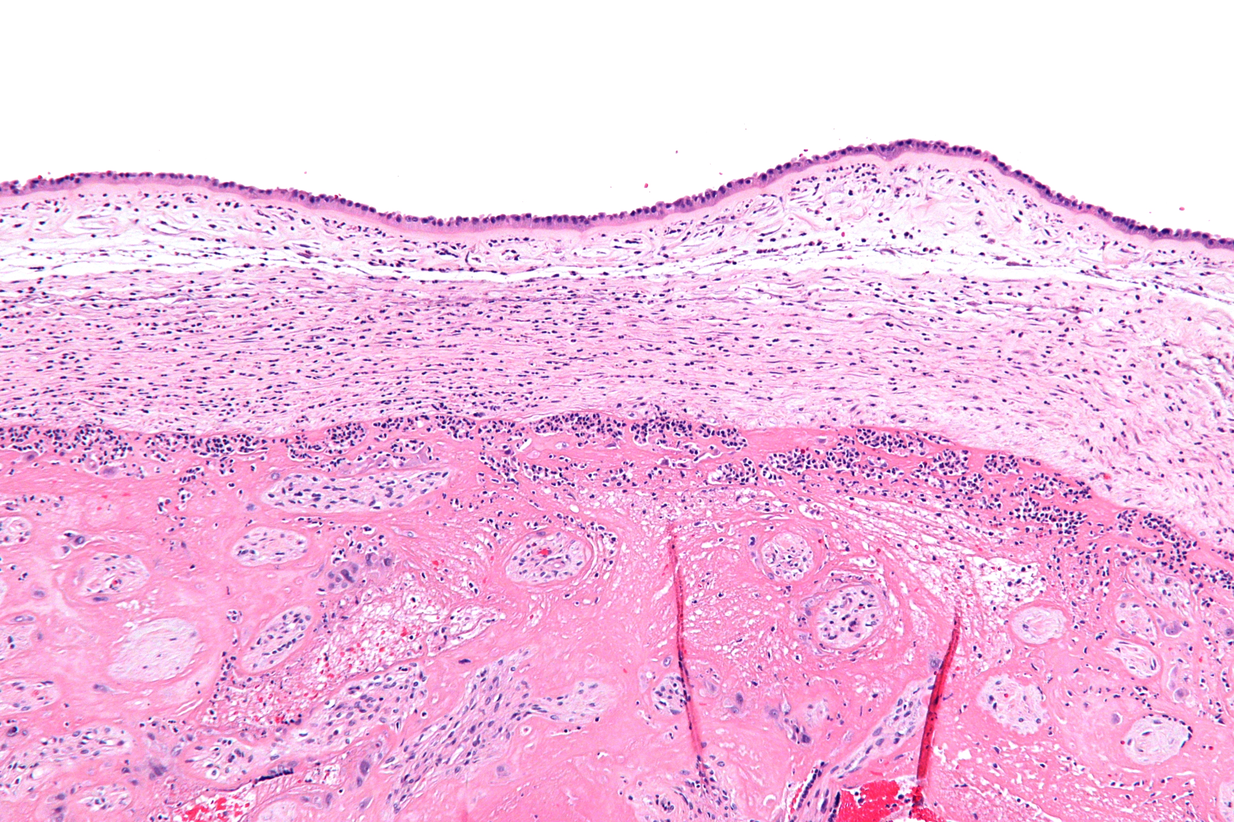
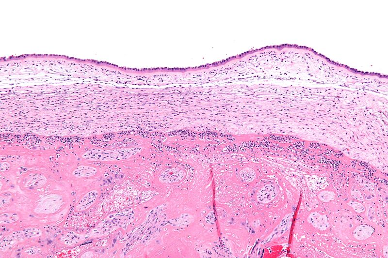
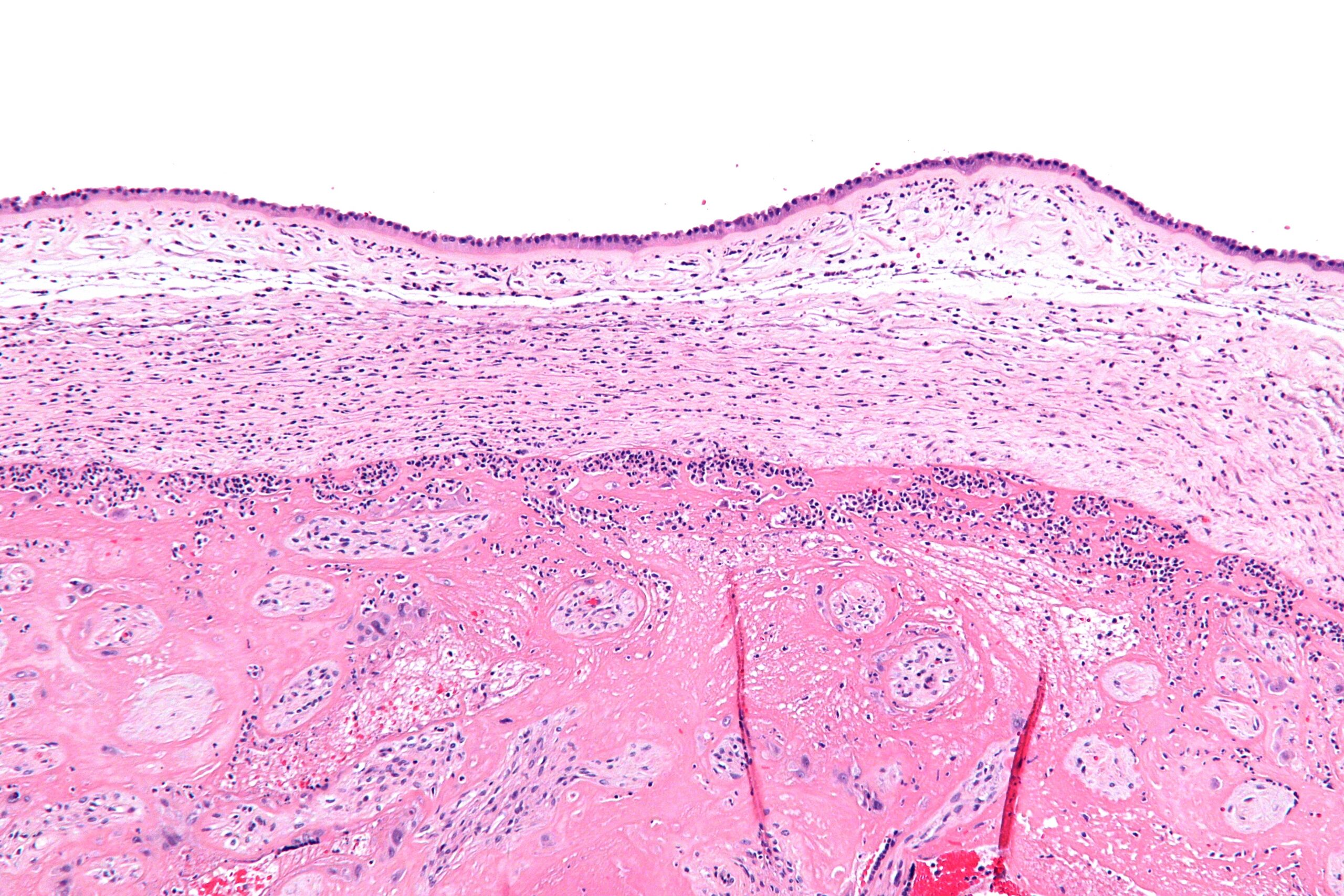
| Description | Intermediate magnification micrograph chorioamnionitis. H&E stain.
The amnion is seen at the very top of the image and composed of a simple cuboidal epithelium and a layer of eosinophilic (pink) connective tissue. It has a few scattered neutrophils - which makes the diagnosis of chorioamnionitis. Below the amnion is a cleft and then the chorion, which also has neutrophils. The fetus (baby) - not shown - would be below the image (and surrounded by amnionic fluid). The pattern of inflammation seen in this image is typical of chorioamnionitis; most chorioamnionitis results from an ascending infection, i.e. the microorganisms ascend from the vagina/cervix. Related images
Low mag.
Intermed mag.
High mag.
Very high mag.
|
| Source | Wikimedia Commons file page |
| Author | Nephron |
| Permission | See original Commons license details. |
Licensing[edit]
Creative Commons Attribution-ShareAlike 3.0 Unported (CC BY-SA 3.0)
This file is licensed under the Creative Commons Attribution-ShareAlike 3.0 license.
Official license: CC BY-SA 3.0
License page: CC BY-SA 3.0
Original attribution and file history: Wikimedia Commons
File history
Click on a date/time to view the file as it appeared at that time.
| Date/Time | Thumbnail | Dimensions | User | Comment | |
|---|---|---|---|---|---|
| current | 03:24, 5 June 2026 | | 4,272 × 2,848 (4.62 MB) | Maintenance script (talk | contribs) | == Summary == Importing file |
You cannot overwrite this file.
File usage
The following 2 pages use this file:
