File:Depth Coded Phalloidin Stained Actin Filaments Cancer Cell.png
Original file (3,176 × 2,472 pixels, file size: 9.86 MB, MIME type: image/png)
Summary[edit]
| Summary | |
|---|---|
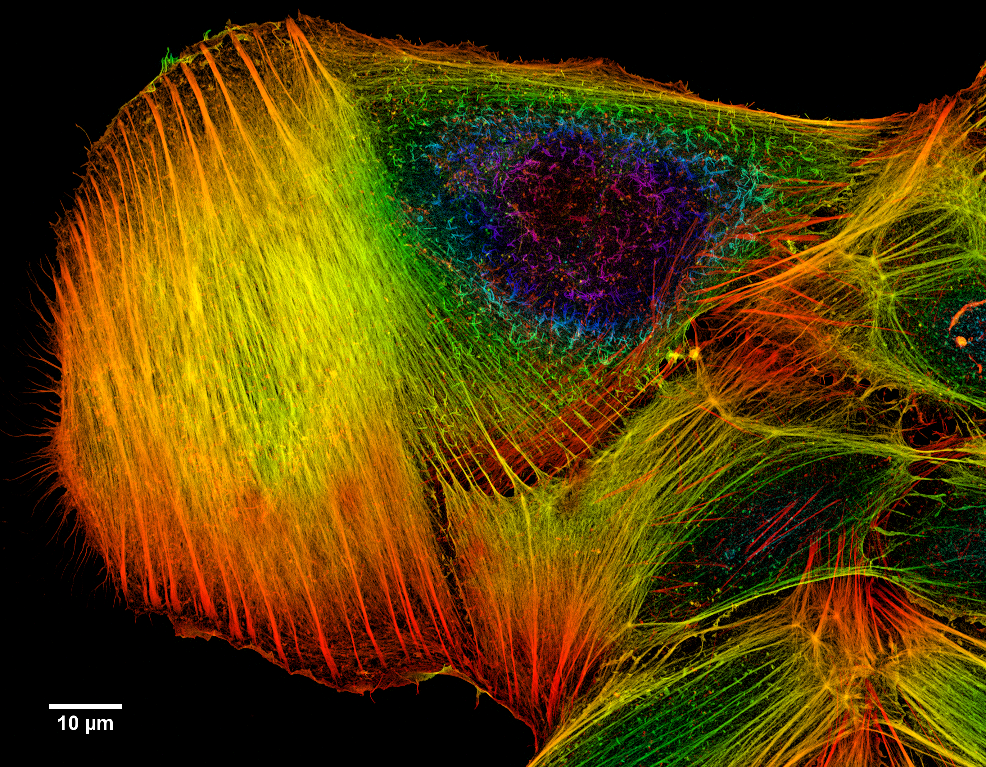
| Description | A z-projection of an osteosarcoma cell, stained with phalloidin to visualise actin filaments. The image was taken on a confocal microscope, and the subsequent deconvolution was done using an experimentally derived point spread function.
Image Parameters Microscope: Zeiss LSM 780 Confocal Microscope Pinhole [m]: 23 µm Total Size-Width: 133.69 µm Total Size-Height: 134.91 µm Total Size-Depth: 6.48 µm Voxel-Width: 42.0 nm (pixel size) Voxel-Height: 42.0 nm (pixel size) Voxel-Depth: 80.0 nm Resolution: 23.5938 pixels per micron Zoom: 1.0 Objective: Plan-Apochromat 63x/1.40 Oil DIC Numerical aperture: 1.40 |
| Source | Wikimedia Commons file page |
| Author | Howard Vindin |
| Permission | See original Commons license details. |
Licensing[edit]
Creative Commons Attribution-ShareAlike 4.0 International (CC BY-SA 4.0)
This file is licensed under the Creative Commons Attribution-ShareAlike 4.0 International license.
You are free to:
- Share — copy and redistribute the material.
- Adapt — remix, transform, and build upon the material.
Under the following conditions:
- Attribution — appropriate credit must be given.
- ShareAlike — derivative works must be distributed under the same license.
Official license: CC BY-SA 4.0
License page: CC BY-SA 4.0
Original attribution and file history: Wikimedia Commons
File history
Click on a date/time to view the file as it appeared at that time.
| Date/Time | Thumbnail | Dimensions | User | Comment | |
|---|---|---|---|---|---|
| current | 03:32, 5 June 2026 | | 3,176 × 2,472 (9.86 MB) | Maintenance script (talk | contribs) | == Summary == Importing file |
You cannot overwrite this file.
File usage
The following file is a duplicate of this file (more details):
- File:Depth Coded Phalloidin Stained Actin Filaments Cancer Cell.png from Wikimedia Commons
The following 3 pages use this file:
