File:Enterobacteria phage T2 transmission electron micrograph.jpg
From WikiMD's WELLNESSPEDIA
Size of this preview: 600 × 600 pixels. Other resolution: 2,021 × 2,021 pixels.
Original file (2,021 × 2,021 pixels, file size: 1.1 MB, MIME type: image/jpeg)
Summary[edit]
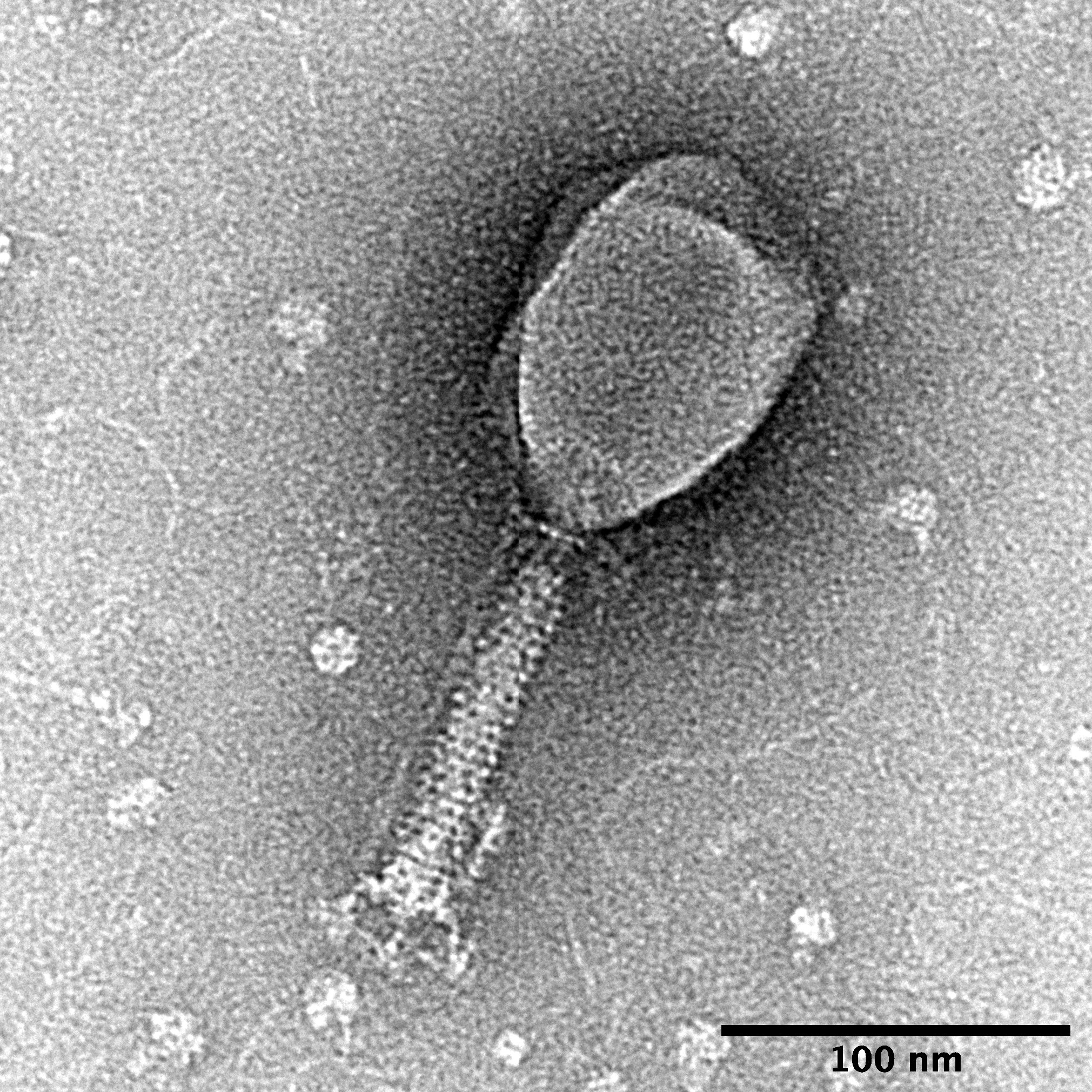
| Summary | |
|---|---|
| Description | T2 phage was prepared from Escherichia coli lysate by PEG precipitation then negatively stained with uranyl formate. Micrograph was taken with an FEI Tecnai transmission electron microscope operating at 120 keV. Cellular debris, nucleic acid, and possibly dissociated tail fibres are visible around the phage. Scale bar, 100 nm. |
| Source | Wikimedia Commons file page |
| Author | SnaxMikn |
| Permission | See original Commons license details. |
Licensing[edit]
Creative Commons Attribution-ShareAlike 4.0 International (CC BY-SA 4.0)
This file is licensed under the Creative Commons Attribution-ShareAlike 4.0 International license.
You are free to:
- Share — copy and redistribute the material.
- Adapt — remix, transform, and build upon the material.
Under the following conditions:
- Attribution — appropriate credit must be given.
- ShareAlike — derivative works must be distributed under the same license.
Official license: CC BY-SA 4.0
Original attribution and file history: Wikimedia Commons
File history
Click on a date/time to view the file as it appeared at that time.
| Date/Time | Thumbnail | Dimensions | User | Comment | |
|---|---|---|---|---|---|
| current | 12:50, 29 May 2026 | | 2,021 × 2,021 (1.1 MB) | Maintenance script (talk | contribs) | == Summary == Importing file |
You cannot overwrite this file.
File usage
The following file is a duplicate of this file (more details):
- File:Enterobacteria phage T2 transmission electron micrograph.jpg from Wikimedia Commons
The following 2 pages use this file:
