File:Fundus photograph of normal right eye.jpg
Original file (1,411 × 1,411 pixels, file size: 248 KB, MIME type: image/jpeg)
Summary[edit]
| Summary | |
|---|---|
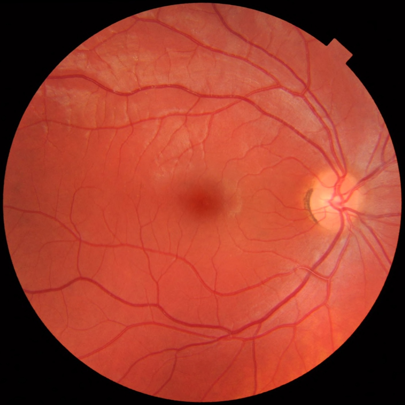
| Description | Fundus photograph of the right eye, showing a fundus with no sign of disease or pathology. It is seen from front so that left in each image is to the person's right. The gaze is into the camera, so the macula is in the center of the image, and the optic disk is located towards the nose (right in image). The optic disk has some pigmentation at the perimeter of the lateral side, which is considered non-pathological.
Veins are darker and slightly wider than corresponding arteries. Major nerve pathways are seen as white striped patterns radiating from the optic disk. In addition, there are also lighter areas close to larger vessels seen mainly at upper left in the image (person's upper right), which is regarded as a normal finding in younger people. Photo is taken at Gävle Hospital in Sweden in 2012 on a healthy 25-year old male volunteer. |
| Source | Wikimedia Commons file page |
| Author | Mikael Häggström.
When using this image in external works, it may be cited as: Häggström, Mikael (2014). "Medical gallery of Mikael Häggström 2014". WikiJournal of Medicine 1 (2). DOI:10.15347/wjm/2014.008. ISSN 2002-4436. Public Domain. or By Mikael Häggström, used with permission. |
| Permission | See original Commons license details. |
Licensing[edit]
License: CC0
License page: CC0
Original attribution and file history: Wikimedia Commons
File history
Click on a date/time to view the file as it appeared at that time.
| Date/Time | Thumbnail | Dimensions | User | Comment | |
|---|---|---|---|---|---|
| current | 12:50, 29 May 2026 | | 1,411 × 1,411 (248 KB) | Maintenance script (talk | contribs) | == Summary == Importing file |
You cannot overwrite this file.
File usage
The following 3 pages use this file:
