File:Gray1110-1.png

Original file (1,301 × 1,305 pixels, file size: 405 KB, MIME type: image/png)
Summary[edit]
| Summary | |
|---|---|
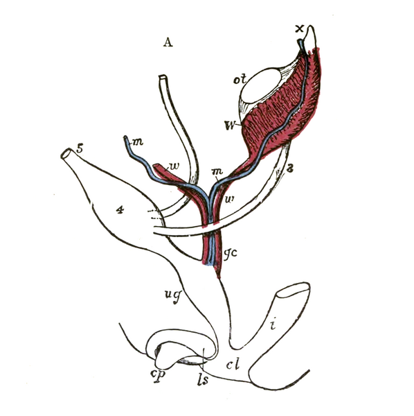
| Description | Diagrams to show the development of male and female generative organs from a common type. (Allen Thomson.) :A.—Diagram of the primitive urogenital organs in the embryo previous to sexual distinction. The common genital cord is labeled with gc.
3. Ureter 4. Urinary bladder 5. Urachus cl. Cloaca cp. Elevation which becomes clitoris or penis i. Lower part of the intestine ls. Fold of integument from which the labia majora or scrotum are formed m, m. Right and left paramesonephric ducts uniting together and running with the mesonephric ducts in gc, the common genital cord ot. The gonadal ridge from which either the ovary or testis is formed ug. Sinus urogenitalis W. Left Wolffian body w, w. Right and left mesonephric ducts.]] |
| Source | Wikimedia Commons file page |
| Author | Henry Vandyke Carter |
| Permission | See original Commons license details. |
Licensing[edit]
Public Domain
This file is in the public domain and may be used without restriction.
Please see the linked source page for the original file history, attribution information, and licensing details.
Original attribution and file history: Wikimedia Commons
File history
Click on a date/time to view the file as it appeared at that time.
| Date/Time | Thumbnail | Dimensions | User | Comment | |
|---|---|---|---|---|---|
| current | 12:49, 29 May 2026 | | 1,301 × 1,305 (405 KB) | Maintenance script (talk | contribs) | == Summary == Importing file |
You cannot overwrite this file.
File usage
The following 2 pages use this file:
