File:HCR-FISH visualization of collagen expression in P. waltl.jpg
Original file (2,048 × 3,624 pixels, file size: 3.06 MB, MIME type: image/jpeg)
Summary[edit]
| Summary | |
|---|---|
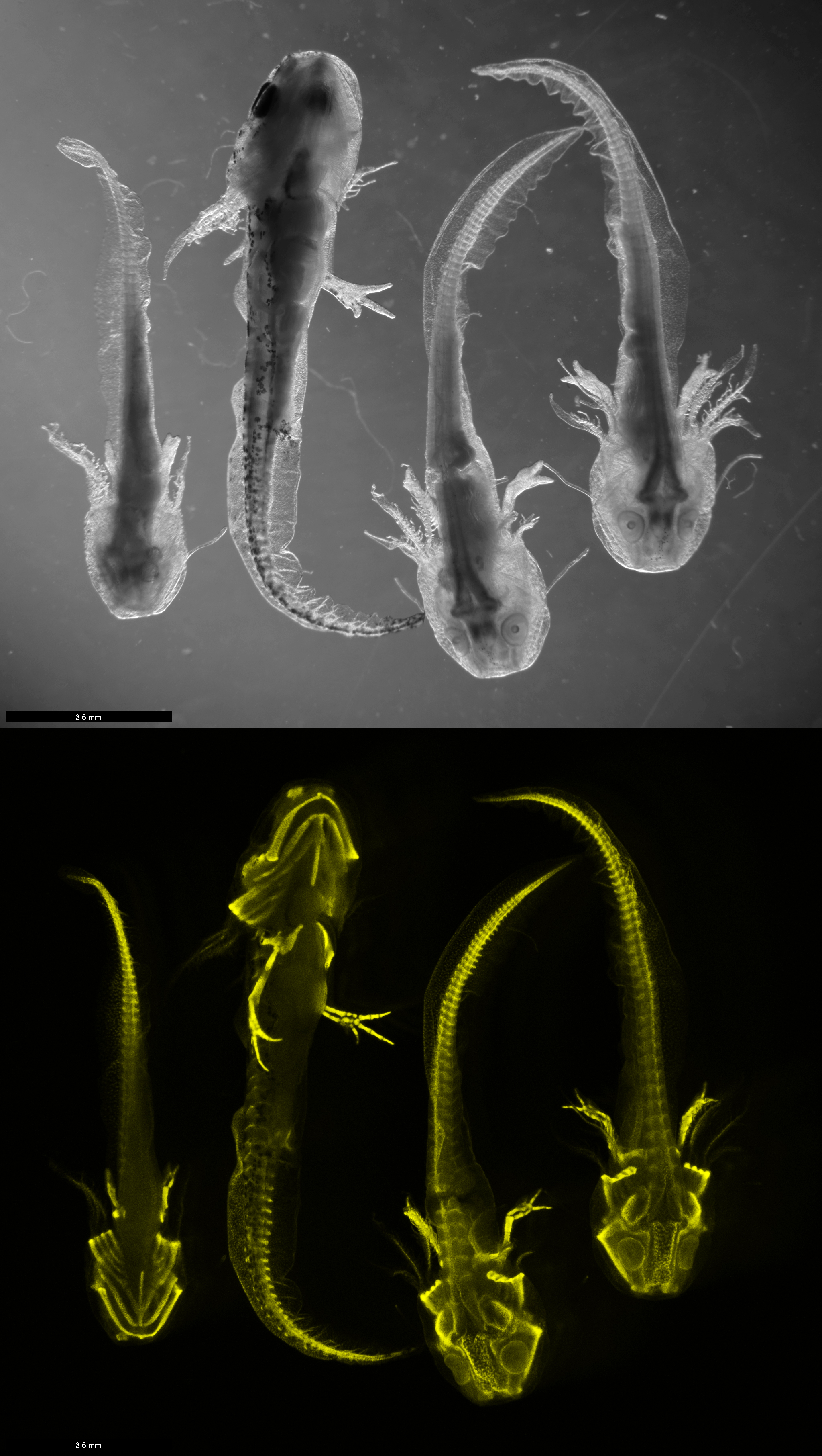
| Description | Top: Black and white bright-field microscopic view of four salamanders (Pleurodeles waltl) at developmental stages 35-37. Specimens 1 and 2 are captured ventrally, whereas specimens 3 and 4 are imaged dorsally. Among these, specimens 1, 3, and 4 are leucistic mutants, characterized by a knocked-out pigmentation gene, while specimen 2 is a wild-type, exhibiting no genetic mutations.
Bottom: The bottom panel illustrates the collagen expression patterns (CO2A1 gene) in these same salamanders, employing Hybridization Chain Reaction RNA Fluorescence In Situ Hybridization (HCR RNA-FISH) technology. The fluorescent visualization predominantly highlights the skeletal areas where collagen is abundantly present. Nonetheless, collagen expression is also observable in other regions, including the gills and on a distinct patch on top of the head. The image was captured by Lennart Rikk from the Regenerative Immunology Lab (https://leighnd.github.io/) using Leica M205 FCA microscope. |
| Source | Wikimedia Commons file page |
| Author | Yodalr |
| Permission | See original Commons license details. |
Licensing[edit]
Creative Commons Attribution 4.0 International (CC BY 4.0)
This file is licensed under the Creative Commons Attribution 4.0 International license.
You may share and adapt the material provided appropriate attribution is given.
Official license: CC BY 4.0
License page: CC BY 4.0
Original attribution and file history: Wikimedia Commons
File history
Click on a date/time to view the file as it appeared at that time.
| Date/Time | Thumbnail | Dimensions | User | Comment | |
|---|---|---|---|---|---|
| current | 03:20, 5 June 2026 | | 2,048 × 3,624 (3.06 MB) | Maintenance script (talk | contribs) | == Summary == Importing file |
You cannot overwrite this file.
File usage
The following 2 pages use this file:
