File:Hammerhead ribozyme ribbons.png
From WikiMD's WELLNESSPEDIA
Size of this preview: 471 × 599 pixels. Other resolution: 637 × 810 pixels.
Original file (637 × 810 pixels, file size: 223 KB, MIME type: image/png)
Summary[edit]
| Summary | |
|---|---|
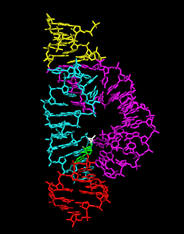
| Description | Sticks bond diagram of full-length hammerhead ribozyme, as determined from the 2.2 Å crystal structure. The figure was created with PyMOL Stem I is in magenta and yellow, stem II is cyan, and stem III is red. The cleavage site nucleotide, C17, is shown in green, and the scissile phosphate is white. |
| Source | Wikimedia Commons file page |
| Author | Wgscott |
| Permission | See original Commons license details. |
Licensing[edit]
Creative Commons Attribution-ShareAlike 3.0 Unported (CC BY-SA 3.0)
This file is licensed under the Creative Commons Attribution-ShareAlike 3.0 license.
Official license: CC BY-SA 3.0
License page: CC BY-SA 3.0
Original attribution and file history: Wikimedia Commons
File history
Click on a date/time to view the file as it appeared at that time.
| Date/Time | Thumbnail | Dimensions | User | Comment | |
|---|---|---|---|---|---|
| current | 03:22, 5 June 2026 | | 637 × 810 (223 KB) | Maintenance script (talk | contribs) | == Summary == Importing file |
You cannot overwrite this file.
File usage
The following 2 pages use this file:
