File:Hexokinase induced fit.svg
From WikiMD's WELLNESSPEDIA

Size of this PNG preview of this SVG file: 537 × 599 pixels. Other resolution: 1,836 × 2,048 pixels.
Original file (SVG file, nominally 1,500 × 1,673 pixels, file size: 1.3 MB)
Summary[edit]
| Summary | |
|---|---|
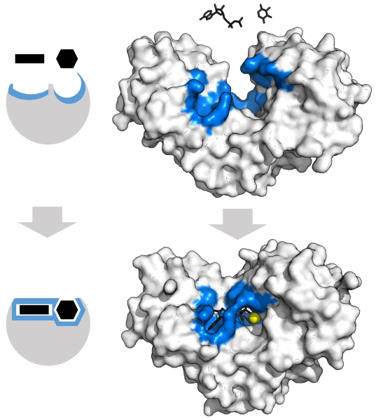
| Description | Enzyme changes shape by induced fit upon substrate binding to form enzyme-substrate complex. Hexokinase has a large induced fit motion that closes over the substrates adenosine triphosphate and xylose. Binding sites in blue, substrates in black and Mg2+ cofactor in yellow. (PDB: 2E2N, 2E2Q) |
| Source | Wikimedia Commons |
| Author | Thomas Shafee |
| Permission | See Commons |
Licensing[edit]
Creative Commons Attribution 4.0 International (CC BY 4.0)
This file is licensed under the Creative Commons Attribution 4.0 International license.
You may share and adapt the material provided appropriate attribution is given.
Official license: CC BY 4.0
Original attribution and file history: Wikimedia Commons
File history
Click on a date/time to view the file as it appeared at that time.
| Date/Time | Thumbnail | Dimensions | User | Comment | |
|---|---|---|---|---|---|
| current | 01:21, 2 June 2026 | | 1,500 × 1,673 (1.3 MB) | Maintenance script (talk | contribs) | == Summary == Importing file |
You cannot overwrite this file.
File usage
The following file is a duplicate of this file (more details):
- File:Hexokinase induced fit.svg from Wikimedia Commons
The following 2 pages use this file:
