File:Histopathology of a bile duct hamartoma, high magnification.jpg
From WikiMD's WELLNESSPEDIA
Size of this preview: 800 × 598 pixels. Other resolution: 2,048 × 1,532 pixels.
Original file (2,048 × 1,532 pixels, file size: 476 KB, MIME type: image/jpeg)
Summary[edit]
| Summary | |
|---|---|
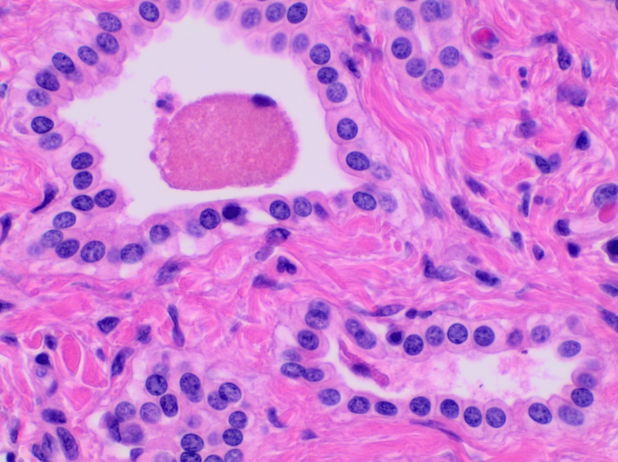
| Description | Histopathology of a bile duct hamartoma, high magnification, H&E stain. It shows typical features of bile duct hamartoma:[1] - Small to medium sized, irregularly shaped bile ducts lined by bland cuboidal epithelium (may also be flattened).- Prominent intervening collagenous stroma. - Bile ducts containing eosinophilic debris (may also contain inspissated bile)
Reference:
|
| Source | Wikimedia Commons file page |
| Author | Mikael Häggström, M.D. Author info - Reusing images- Conflicts of interest: None Mikael Häggström, M.D.Consent note: Consent from the patient or patient's relatives is regarded as redundant, because of absence of identifiable features (List of HIPAA identifiers) in the media and case information (See also HIPAA case reports guidance). |
| Permission | See original Commons license details. |
Licensing[edit]
License: CC0
License page: CC0
Original attribution and file history: Wikimedia Commons
File history
Click on a date/time to view the file as it appeared at that time.
| Date/Time | Thumbnail | Dimensions | User | Comment | |
|---|---|---|---|---|---|
| current | 03:36, 5 June 2026 | | 2,048 × 1,532 (476 KB) | Maintenance script (talk | contribs) | == Summary == Importing file |
You cannot overwrite this file.
File usage
The following file is a duplicate of this file (more details):
- File:Histopathology of a bile duct hamartoma, high magnification.jpg from Wikimedia Commons
The following 2 pages use this file:
