File:Histopathology of adenocarcinoma.png
From WikiMD's WELLNESSPEDIA
Size of this preview: 701 × 600 pixels. Other resolution: 1,532 × 1,311 pixels.
Original file (1,532 × 1,311 pixels, file size: 2.28 MB, MIME type: image/png)
Summary[edit]
| Summary | |
|---|---|
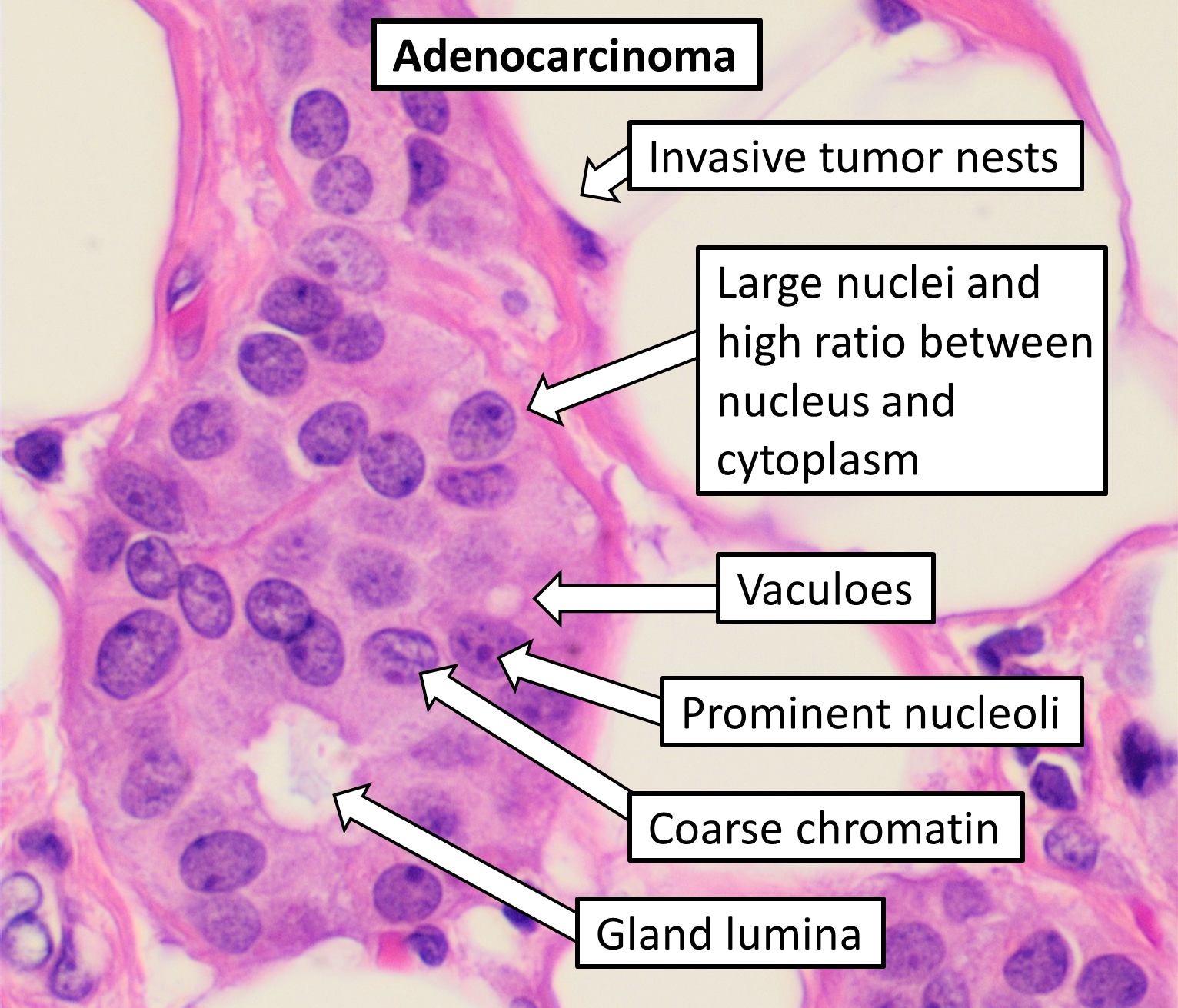
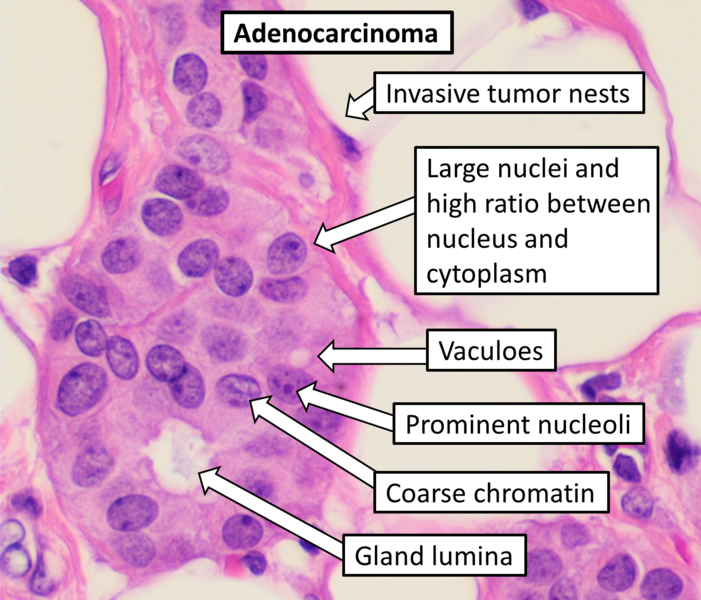
| Description | Histopathology of adenocarcinoma, H&E stain, with typical features. This case is an invasive carcinoma of no special type of the breast. In this case, invasion is supported by presence within fatty tissue (which, however, may also occur in benign conditions such as microglandular adenosis). The large size of the nuclei can be appreciated by comparing to the benign dark stromal cell by the left image border. In reality, the visual features vary substantially, both by subtypes of adenocarcinoma as well as between individual cases. |
| Source | Wikimedia Commons file page |
| Author | Mikael Häggström, M.D. Author info - Reusing images- Conflicts of interest: None Mikael Häggström, M.D.Consent note: Consent from the patient or patient's relatives is regarded as redundant, because of absence of identifiable features (List of HIPAA identifiers) in the media and case information (See also HIPAA case reports guidance). |
| Permission | See original Commons license details. |
Licensing[edit]
License: CC0
License page: CC0
Original attribution and file history: Wikimedia Commons
File history
Click on a date/time to view the file as it appeared at that time.
| Date/Time | Thumbnail | Dimensions | User | Comment | |
|---|---|---|---|---|---|
| current | 03:18, 5 June 2026 | | 1,532 × 1,311 (2.28 MB) | Maintenance script (talk | contribs) | == Summary == Importing file |
You cannot overwrite this file.
File usage
The following file is a duplicate of this file (more details):
- File:Histopathology of adenocarcinoma.png from Wikimedia Commons
The following 2 pages use this file:
