File:Histopathology of carcinosarcoma, annotated.png
From WikiMD's WELLNESSPEDIA
Size of this preview: 800 × 598 pixels. Other resolution: 2,048 × 1,532 pixels.
Original file (2,048 × 1,532 pixels, file size: 7.13 MB, MIME type: image/png)
Summary[edit]
| Summary | |
|---|---|
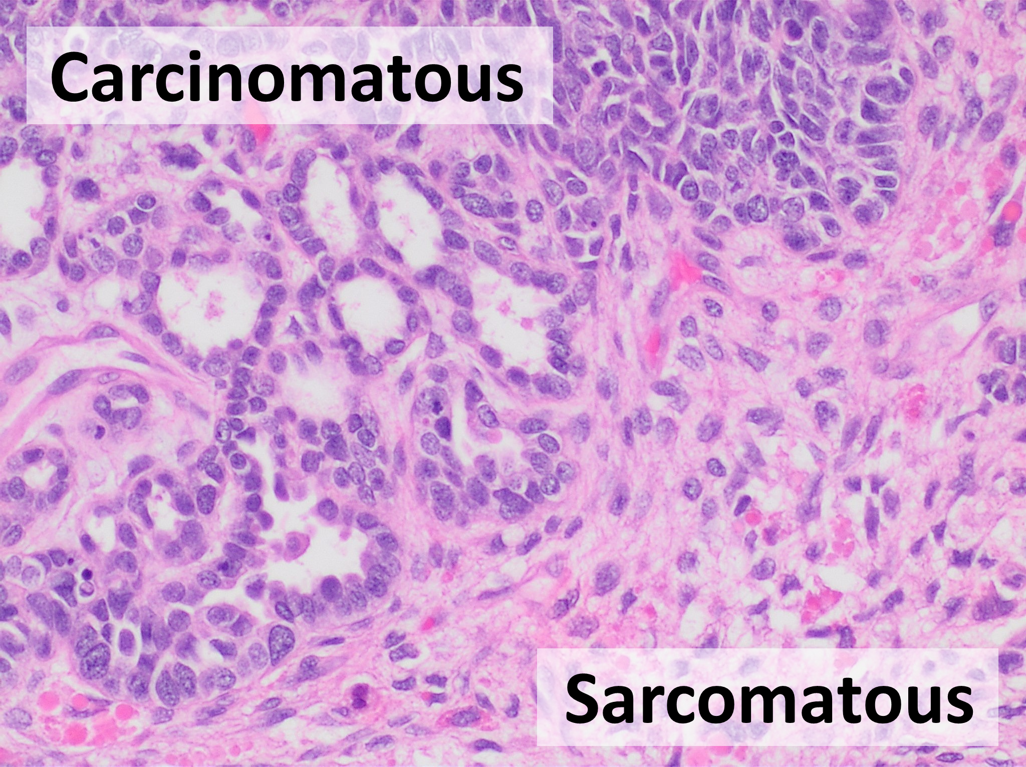
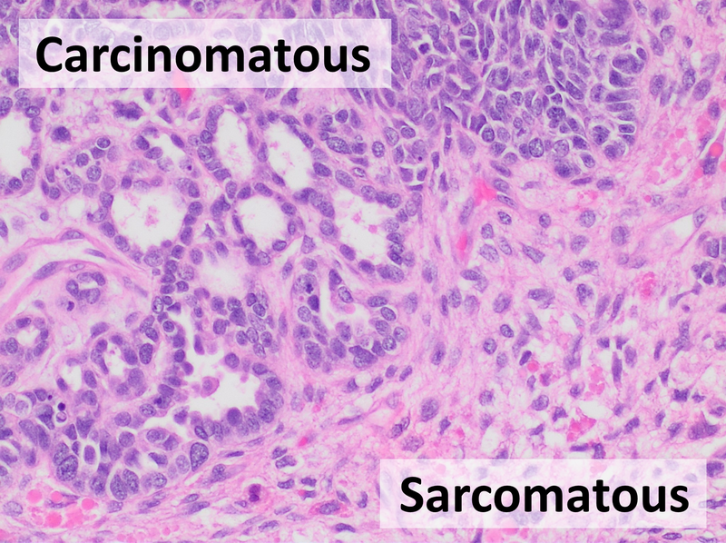
| Description | Histopathology of carcinosarcoma from an ovary, H&E stain, showing:- carcinomatous element at top left, such as having gland formations- sarcomatous element at bottom right, such as having more spindle-shaped nuclei and abundant extracellular matrix. |
| Source | Wikimedia Commons file page |
| Author | Mikael Häggström, M.D. Author info - Reusing images- Conflicts of interest: None Mikael Häggström, M.D.Consent note: Consent from the patient or patient's relatives is regarded as redundant, because of absence of identifiable features (List of HIPAA identifiers) in the media and case information (See also HIPAA case reports guidance). |
| Permission | See original Commons license details. |
Licensing[edit]
License: CC0
License page: CC0
Original attribution and file history: Wikimedia Commons
File history
Click on a date/time to view the file as it appeared at that time.
| Date/Time | Thumbnail | Dimensions | User | Comment | |
|---|---|---|---|---|---|
| current | 03:18, 5 June 2026 | | 2,048 × 1,532 (7.13 MB) | Maintenance script (talk | contribs) | == Summary == Importing file |
You cannot overwrite this file.
File usage
The following 2 pages use this file:
