File:Histopathology of dense fibrous scar replacing myocyte loss in myocardial infarction.jpg
From WikiMD's WELLNESSPEDIA
No higher resolution available.
Histopathology_of_dense_fibrous_scar_replacing_myocyte_loss_in_myocardial_infarction.jpg (751 × 566 pixels, file size: 211 KB, MIME type: image/jpeg)
Summary[edit]
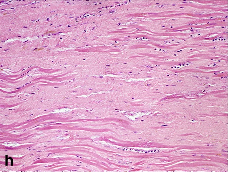
| Summary | |
|---|---|
| Description | Histological features of MI at different stages, without reperfusion; (a) myofiber waviness (b) interstitial oedema(c) hypereosinophilia and coagulative necrosis of cardiomyocytes (d) heavy granulocyte infiltration with karyorrhexis (e) macrophages and lymphocyte infiltration with early removal of necrotic debris (f) granulation tissue with formation of microvessels (g) fibroblast proliferation and early collagen deposition (h) dense fibrous scar replacing myocyte loss All sections are stained with haematoxylin and eosin. |
| Source | Wikimedia Commons |
| Author | Katarzyna Michaud, Cristina Basso, Giulia d’Amati, Carla Giordano, Ivana Kholová, Stephen D. Preston, Stefania Rizzo, Sara Sabatasso, Mary N. Sheppard, Aryan Vink, Allard C. van der Wal & on behalf of the Association for European Cardiovascular Pathology (AECVP) |
| Permission | See Commons |
Licensing[edit]
Creative Commons Attribution 3.0 Unported (CC BY 3.0)
This file is licensed under the Creative Commons Attribution 3.0 license.
Official license: CC BY 3.0
Original attribution and file history: Wikimedia Commons
File history
Click on a date/time to view the file as it appeared at that time.
| Date/Time | Thumbnail | Dimensions | User | Comment | |
|---|---|---|---|---|---|
| current | 01:21, 2 June 2026 | | 751 × 566 (211 KB) | Maintenance script (talk | contribs) | == Summary == Importing file |
You cannot overwrite this file.
File usage
The following file is a duplicate of this file (more details):
The following 2 pages use this file:
