File:Histopathology of invasive low-grade serous carcinoma of ovary.png
From WikiMD's WELLNESSPEDIA
Size of this preview: 800 × 546 pixels. Other resolutions: 2,560 × 1,746 pixels | 3,209 × 2,189 pixels.
Original file (3,209 × 2,189 pixels, file size: 9 MB, MIME type: image/png)
Summary[edit]
| Summary | |
|---|---|
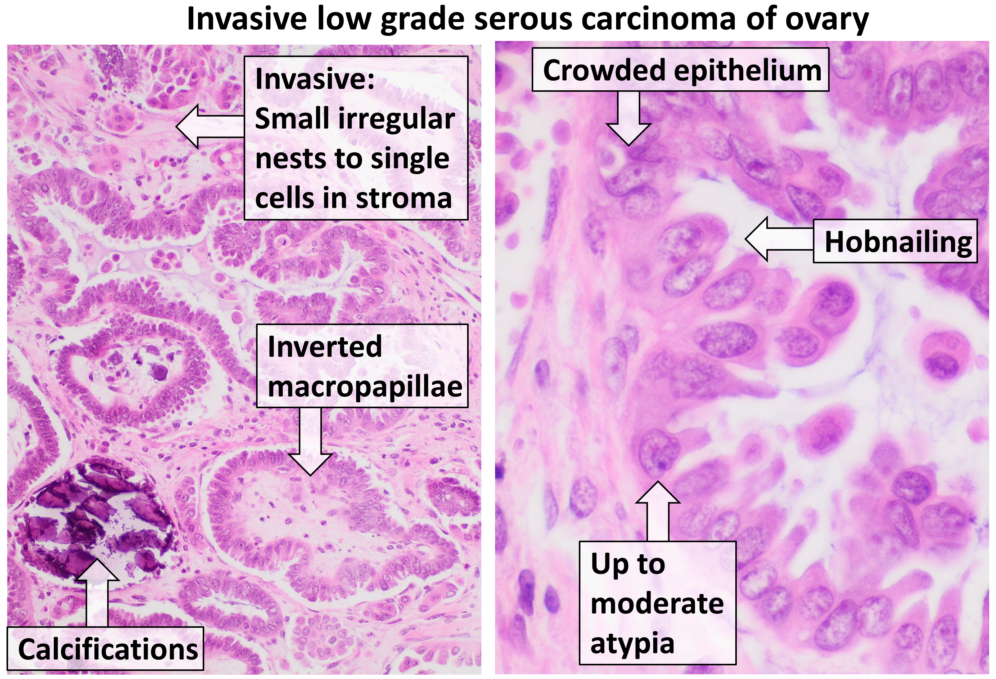
| Description | Histopathology of invasive low-grade serous carcinoma of ovary with typical features. H&E stain. The left image shows lower magnification, including inverted macropapillae which are with broad fibrovascular cores surrounded by clear (white) clefts. Invasion (characterized by small irregular nests to single cells) should be over 5 mm in size to distinguish it from a borderline serous tumor. Calcifications often form psammoma bodies. Right picture shows higher magnification, including hobnailing which is individual cells protruding into the lumen of glands. Cells may have up to moderate atypia: They may have conspicuous nucleoli, and up to 3x variation in nuclear sizes compared to each other. More atypical features indicate a high-grade serous carcinoma. Reference for findings: Erna Forgó, M.D., Teri A. Longacre, M.D.. Low grade serous carcinoma. Pathology Outlines. Last staff update: 23 July 2020 |
| Source | Wikimedia Commons file page |
| Author | Mikael Häggström, M.D. Author info - Reusing images- Conflicts of interest: None Mikael Häggström, M.D.Consent note: Consent from the patient or patient's relatives is regarded as redundant, because of absence of identifiable features (List of HIPAA identifiers) in the media and case information (See also HIPAA case reports guidance). |
| Permission | See original Commons license details. |
Licensing[edit]
License: CC0
License page: CC0
Original attribution and file history: Wikimedia Commons
File history
Click on a date/time to view the file as it appeared at that time.
| Date/Time | Thumbnail | Dimensions | User | Comment | |
|---|---|---|---|---|---|
| current | 22:33, 8 June 2026 | | 3,209 × 2,189 (9 MB) | Maintenance script (talk | contribs) | == Summary == Importing file |
You cannot overwrite this file.
File usage
The following file is a duplicate of this file (more details):
- File:Histopathology of invasive low-grade serous carcinoma of ovary.png from Wikimedia Commons
The following page uses this file:
