File:Histopathology of liposarcoma, annotated.jpg
From WikiMD's WELLNESSPEDIA

Size of this preview: 792 × 600 pixels. Other resolution: 1,945 × 1,473 pixels.
Original file (1,945 × 1,473 pixels, file size: 619 KB, MIME type: image/jpeg)
Summary[edit]
| Summary | |
|---|---|
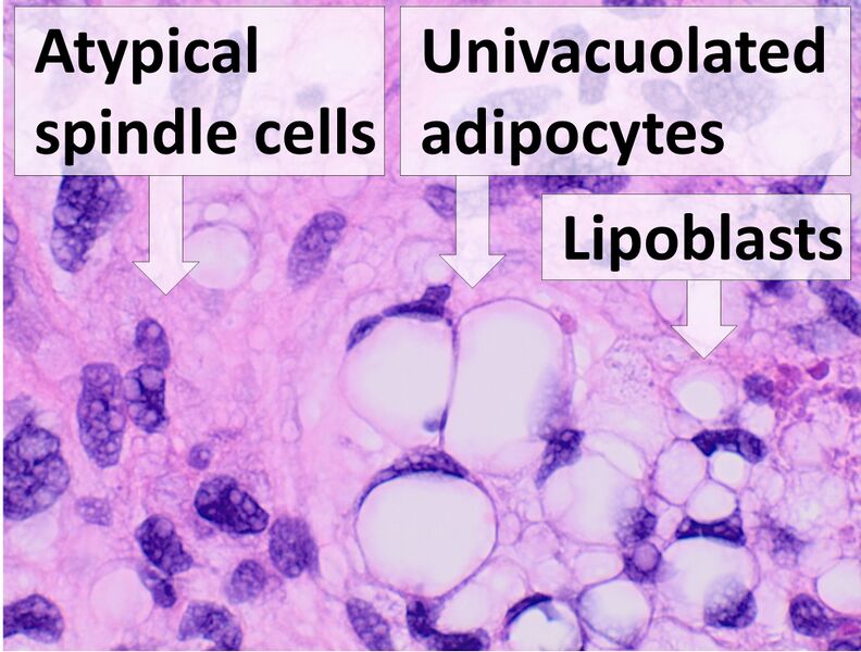
| Description | Histopathology of liposarcoma, H&E stain, with the main features:[1]- Spindle cells with enlarged, hyperchromatic nuclei.- Apparently univacuolated adipocytes (may look normal).- Lipoblasts (multivacuolated), but neither necessary nor sufficient for diagnosis.
This case was likely a myxoid liposarcoma as per immunohistochemistry. Reference:
|
| Source | Wikimedia Commons file page |
| Author | Mikael Häggström, M.D. Author info - Reusing images- Conflicts of interest: None Mikael Häggström, M.D.Consent note: Consent from the patient or patient's relatives is regarded as redundant, because of absence of identifiable features (List of HIPAA identifiers) in the media and case information (See also HIPAA case reports guidance). |
| Permission | See original Commons license details. |
Licensing[edit]
License: CC0
License page: CC0
Original attribution and file history: Wikimedia Commons
File history
Click on a date/time to view the file as it appeared at that time.
| Date/Time | Thumbnail | Dimensions | User | Comment | |
|---|---|---|---|---|---|
| current | 03:30, 5 June 2026 | | 1,945 × 1,473 (619 KB) | Maintenance script (talk | contribs) | == Summary == Importing file |
You cannot overwrite this file.
File usage
The following 2 pages use this file:
