File:Histopathology of seminoma.png
From WikiMD's WELLNESSPEDIA
Size of this preview: 800 × 553 pixels. Other resolutions: 2,560 × 1,769 pixels | 3,071 × 2,122 pixels.
Original file (3,071 × 2,122 pixels, file size: 8.73 MB, MIME type: image/png)
Summary[edit]
| Summary | |
|---|---|
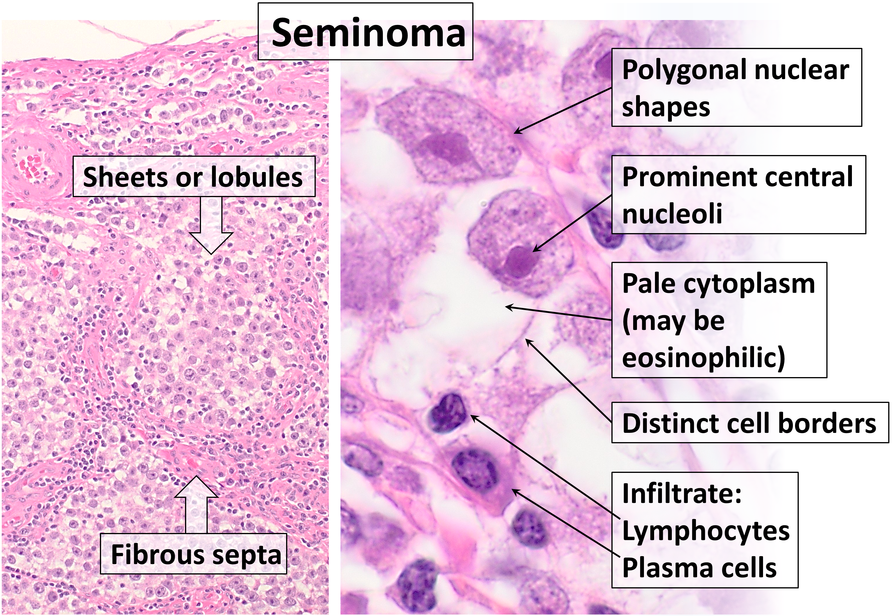
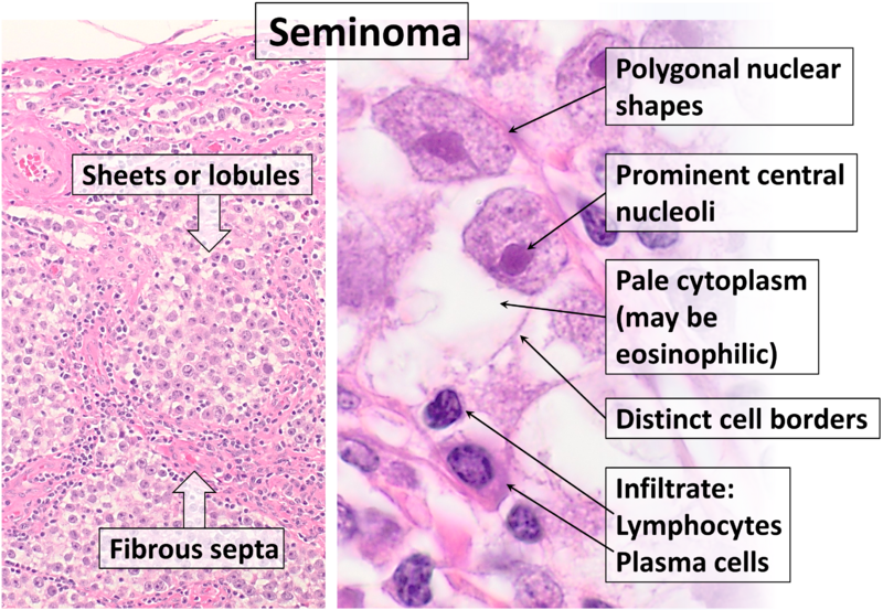
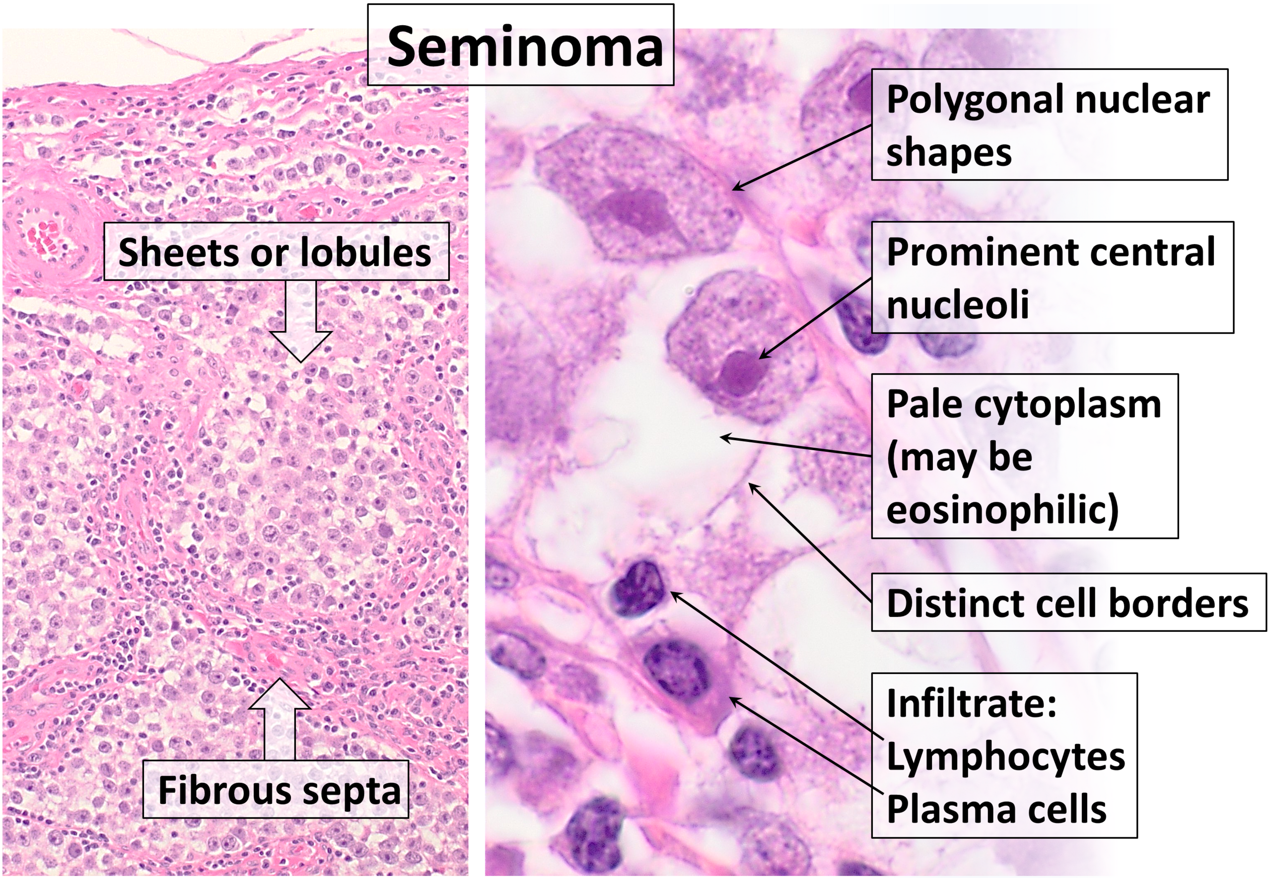
| Description | Histopathology of classical seminoma, with typical features. H&E stain. Reference for findings: Michelle R. Downes, M.D.. Testis & paratestis - Seminoma. Pathology Outlines. Last author update: 7 January 2020. Last staff update: 19 April 2022 |
| Source | Wikimedia Commons file page |
| Author | Mikael Häggström, M.D. Author info - Reusing images- Conflicts of interest: None Mikael Häggström, M.D.Consent note: Consent from the patient or patient's relatives is regarded as redundant, because of absence of identifiable features (List of HIPAA identifiers) in the media and case information (See also HIPAA case reports guidance). |
| Permission | See original Commons license details. |
Licensing[edit]
License: CC0
License page: CC0
Original attribution and file history: Wikimedia Commons
File history
Click on a date/time to view the file as it appeared at that time.
| Date/Time | Thumbnail | Dimensions | User | Comment | |
|---|---|---|---|---|---|
| current | 22:35, 8 June 2026 | | 3,071 × 2,122 (8.73 MB) | Maintenance script (talk | contribs) | == Summary == Importing file |
You cannot overwrite this file.
File usage
The following page uses this file:
