File:Human brainstem-thalamus posterior view description.JPG
Human_brainstem-thalamus_posterior_view_description.JPG (340 × 485 pixels, file size: 23 KB, MIME type: image/jpeg)
Summary[edit]
| Summary | |
|---|---|
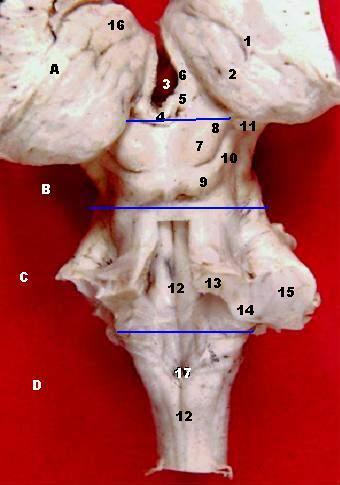
| Description | Human brainstem and thalamus - posterior view
Taenia choroidea (and lateral: Lamina affixa, Stria terminalis) Thalamus, Pulvinar thalami (Ventriculus tertius) Stalk of Glandula pinealis Habenula Stria medullaris Colliculus superior Brachium colliculi superioris Colliculus inferior Brachium colliculi inferioris Corpus geniculatum mediale Sulcus medianus Pedunculus cerebellaris superior Pedunculus cerebellaris inferior Pedunculus cerebellaris medius Tuberculum anterius thalami Obex, Area postrema A: Thalamus, B: Mesencephalon, C: Pons, D: Medulla oblongata On this specimen, the following thalamic structures can be seen: 1. the Epithalamus (Stria Medullaris Thalami, Habenula, & Pineal), 2. the Anterior Nucleus of the dorsal thalamus (Anterior Tubercle) and, 3. the Pulvinar (the large posterior portion of the dorsal thalamus which overhangs the midbrain. The Medulla, Pons & Midbrain are delineated on the posterior surface of the brainstem. NOTE: The 4 Colliculi of the tectum are refered to collectively as the Quadrigeminal Plate. The three Cerebellar Peduncles are shown here as they enter the brainstem on each side. In the Midbrain identify the Superior Colliculus and Inferior Colliculus. Also identify the Brachium of the Superior Colliculus and the Brachium of the Inferior Colliculus which connect with the Lateral Geniculate Body and Medial Geniculate Body, respectively. The cerebellum forms the roof of the 4th ventricle and is connected to the brainstem by 3 pairs of peduncles or pillars (shown on right side of brainstem) . The peduncles are made up of axons entering and leaving the cerebellum. The Inferior Cerebellar peduncle projects from the medulla, the large Middle Cerebellar Peduncle projects from the Pons, and the Superior Cerebellar Peduncle connects with the midbrain.
|
| Source | Wikimedia Commons file page |
| Author | John A Beal, PhD Dep't. of Cellular Biology & Anatomy, Louisiana State University Health Sciences Center Shreveport |
| Permission | See original Commons license details. |
Licensing[edit]
License: CC BY 2.5
License page: CC BY 2.5
Original attribution and file history: Wikimedia Commons
File history
Click on a date/time to view the file as it appeared at that time.
| Date/Time | Thumbnail | Dimensions | User | Comment | |
|---|---|---|---|---|---|
| current | 12:50, 29 May 2026 | | 340 × 485 (23 KB) | Maintenance script (talk | contribs) | == Summary == Importing file |
You cannot overwrite this file.
File usage
The following file is a duplicate of this file (more details):
- File:Human brainstem-thalamus posterior view description.JPG from Wikimedia Commons
The following 4 pages use this file:
