File:Human subventricular zone.jpg
From WikiMD's WELLNESSPEDIA

Size of this preview: 800 × 369 pixels. Other resolution: 1,200 × 553 pixels.
Original file (1,200 × 553 pixels, file size: 166 KB, MIME type: image/jpeg)
Summary[edit]
| Summary | |
|---|---|
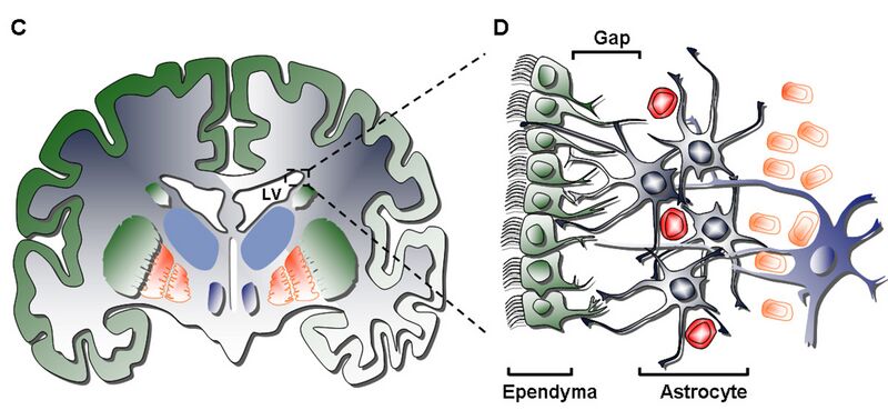
| Description | The anatomy of the neurogenic subventricular zone in the adult rodent and human brain. (A) Sagittal view of a rodent brain showing the sites of neurogenesis in the subventricular zone/olfactory bulb (SVZ/OB) system. (B) Schematic drawing of the composition and cytoarchitecture of the adult rodent SVZ. (C) Coronal view of the adult human brain showing the basal ganglia and lateral ventricles. (D) Schematic drawing depicting the cellular composition and cytoarchitecture of the adult human SVZ, consisting of four layers: Layer I – ependymal cell layer (green), Layer II – hypocellular gap, Layer III – astrocytic ribbon, containing astrocytes and migrating neuroblasts, Layer IV – transitional zone, containing oligodendrocytes and separating the SVZ from the striatum rich in neurons. RMS: rostral migratory stream; DG: dentate gyrus; LV: lateral ventricle. Arias-Carrión International Archives of Medicine 2008 1:2 doi:10.1186/1755-7682-1-2 |
| Source | Wikimedia Commons file page |
| Author | The_anatomy_of_the_neurogenic_subventricular_zone_in_the_adult_rodent_and_human_brain.jpg: Oscar Arias-Carrión derivative work: CopperKettle (talk) |
| Permission | See original Commons license details. |
Licensing[edit]
License: CC BY 2.0
License page: CC BY 2.0
Original attribution and file history: Wikimedia Commons
File history
Click on a date/time to view the file as it appeared at that time.
| Date/Time | Thumbnail | Dimensions | User | Comment | |
|---|---|---|---|---|---|
| current | 03:27, 5 June 2026 | | 1,200 × 553 (166 KB) | Maintenance script (talk | contribs) | == Summary == Importing file |
You cannot overwrite this file.
File usage
The following 2 pages use this file:
