File:Leydig cell tumour2.jpg
Original file (4,272 × 2,848 pixels, file size: 3.41 MB, MIME type: image/jpeg)
Summary[edit]
| Summary | |
|---|---|
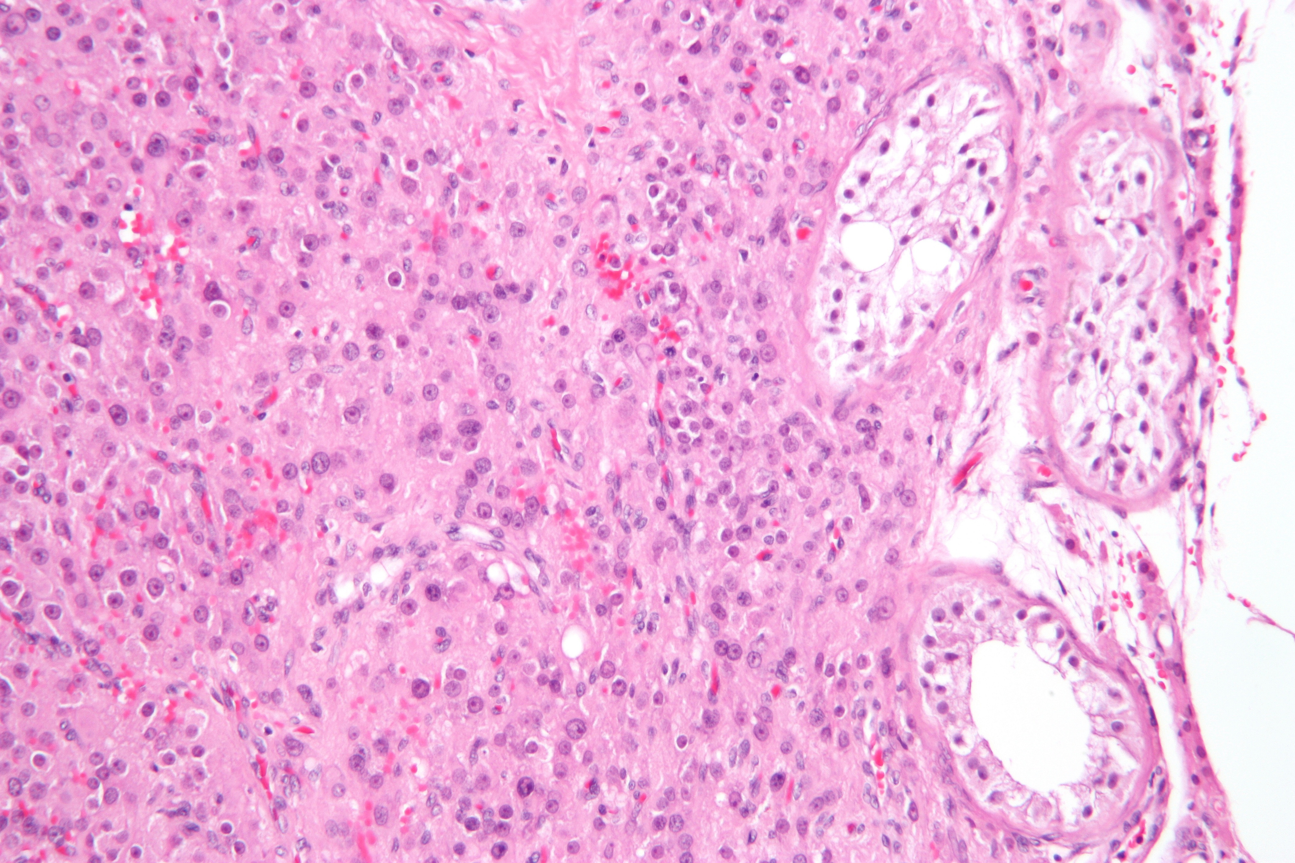
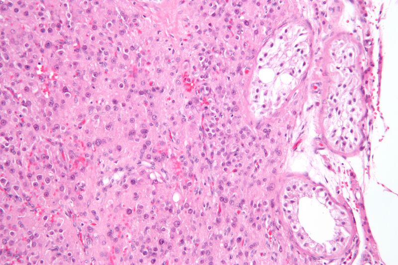
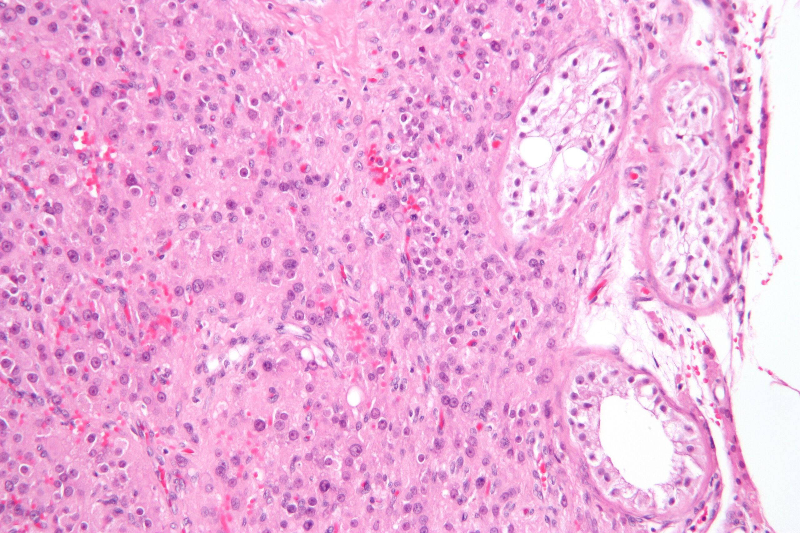
| Description | Intermediate magnification micrograph of a Leydig cell tumour of the testis. H&E stain. Leydig cell tumours fall in the sex cord-stromal tumour subgroup of testicular tumours.
Leydig tumour shown has: cells with moderate nuclear size variation, and cells with: prominent round central nucleoli, and an eosinophilic vacuolated cytoplasm. Classically, Leydig tumours have Reinke crystals, cylindrical crystalloid eosinophilic cytoplasmic bodies; these are not apparent in the micrograph. See also Image:Leydig_cell_tumour1.jpg - low magnification view. Image:Leydig_cell_tumour3.jpg - high magnification view. |
| Source | Wikimedia Commons file page |
| Author | Nephron |
| Permission | See original Commons license details. |
Licensing[edit]
Creative Commons Attribution-ShareAlike 3.0 Unported (CC BY-SA 3.0)
This file is licensed under the Creative Commons Attribution-ShareAlike 3.0 license.
Official license: CC BY-SA 3.0
License page: CC BY-SA 3.0
Original attribution and file history: Wikimedia Commons
File history
Click on a date/time to view the file as it appeared at that time.
| Date/Time | Thumbnail | Dimensions | User | Comment | |
|---|---|---|---|---|---|
| current | 03:30, 5 June 2026 | | 4,272 × 2,848 (3.41 MB) | Maintenance script (talk | contribs) | == Summary == Importing file |
You cannot overwrite this file.
File usage
The following 3 pages use this file:
