File:Lissencephaly.png
From WikiMD's WELLNESSPEDIA
Size of this preview: 521 × 600 pixels. Other resolution: 928 × 1,068 pixels.
Original file (928 × 1,068 pixels, file size: 1.56 MB, MIME type: image/png)
Summary[edit]
| Summary | |
|---|---|
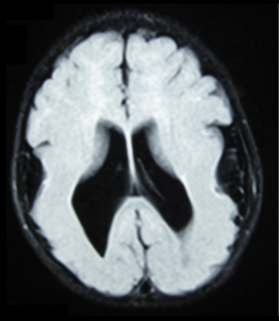
| Description | Brain MRI, T1 weighted, transverse plane, that shows lyssencephaly, manifested as scarce and wide circumvolutions, mostly in the occipital, parietal and temporal lobes. As aggregated findings, there is ventriculomegaly, no true Sylvian cissure, too thick gray matter and ectopic gray matter in the white matter. |
| Source | Wikimedia Commons file page |
| Author | Ralphelg |
| Permission | See original Commons license details. |
Licensing[edit]
Creative Commons Attribution-ShareAlike 3.0 Unported (CC BY-SA 3.0)
This file is licensed under the Creative Commons Attribution-ShareAlike 3.0 license.
Official license: CC BY-SA 3.0
License page: CC BY-SA 3.0
Original attribution and file history: Wikimedia Commons
File history
Click on a date/time to view the file as it appeared at that time.
| Date/Time | Thumbnail | Dimensions | User | Comment | |
|---|---|---|---|---|---|
| current | 03:32, 5 June 2026 | | 928 × 1,068 (1.56 MB) | Maintenance script (talk | contribs) | == Summary == Importing file |
You cannot overwrite this file.
File usage
The following 2 pages use this file:
