File:Macular corneal dystrophy hale colloidal iron stain.JPEG
From WikiMD's WELLNESSPEDIA
No higher resolution available.
Macular_corneal_dystrophy_hale_colloidal_iron_stain.JPEG (500 × 500 pixels, file size: 152 KB, MIME type: image/jpeg)
Summary[edit]
| Summary | |
|---|---|
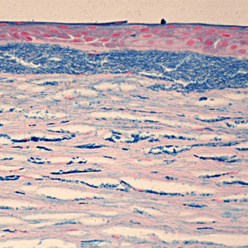
| Description | Macular corneal dystrophy. The abnormalities within the cornea are easily seen within the keratocytes and in a subepithelial extracellular location because they stain prominently with methods that demonstrate glycosaminoglycans. Hale colloidal iron stain. Klintworth Orphanet Journal of Rare Diseases 2009 4:7 doi:10.1186/1750-1172-4-7 |
| Source | Wikimedia Commons file page |
| Author | Klintworth GK. |
| Permission | See original Commons license details. |
Licensing[edit]
License: CC BY 2.0
License page: CC BY 2.0
Original attribution and file history: Wikimedia Commons
File history
Click on a date/time to view the file as it appeared at that time.
| Date/Time | Thumbnail | Dimensions | User | Comment | |
|---|---|---|---|---|---|
| current | 22:29, 8 June 2026 | | 500 × 500 (152 KB) | Maintenance script (talk | contribs) | == Summary == Importing file |
You cannot overwrite this file.
File usage
The following page uses this file:
