File:Mantle cell lymphoma - intermed mag.jpg
Original file (2,848 × 4,272 pixels, file size: 4.12 MB, MIME type: image/jpeg)
Summary[edit]
| Summary | |
|---|---|
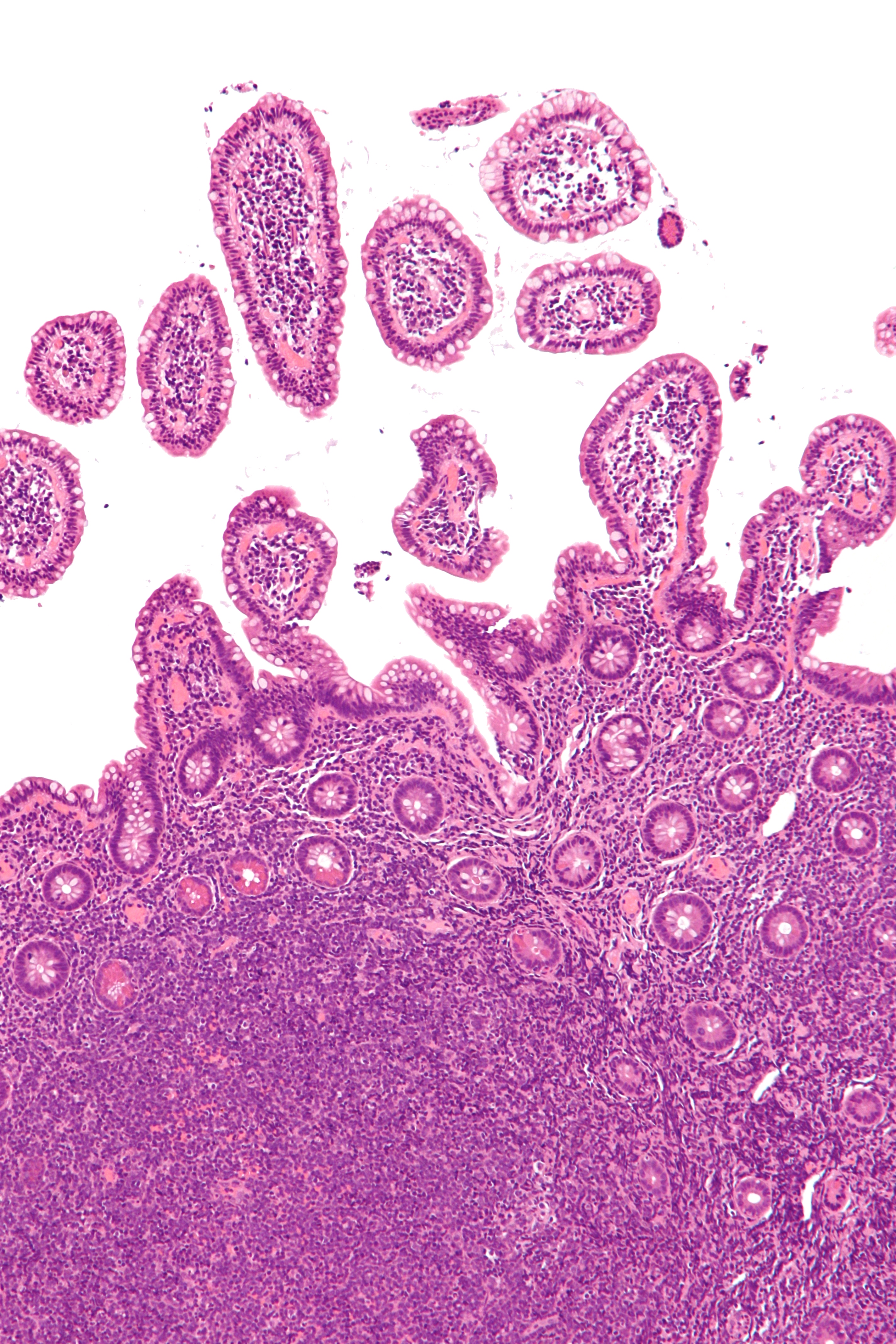
| Description | Intermediate magnification micrograph of mantle cell lymphoma of the terminal ileum. Endoscopic biopsy. H&E stain.
Histomorphologic features: Monomorphic small lymphoid cells less than twice the size of a resting lymphocyte. Abundant mitoses. Sclerosed blood vessels. Scattered epithelioid histiocytes. Immunohistochemical staining: Positive: Cyclin D1, CD5, CD43, CD20, CD45. Negative: CD23. Molecular features: t(11;14)(q13;q32). CCND1/IGHG1 fusion gene.[1] E Related images
Low mag.
Intermed. mag.
Low mag. - cyclin D1.
Intermed. mag. - cyclin D1.
|
| Source | Wikimedia Commons |
| Author | Nephron |
| Permission | See Commons |
Licensing[edit]
Creative Commons Attribution-ShareAlike 3.0 Unported (CC BY-SA 3.0)
This file is licensed under the Creative Commons Attribution-ShareAlike 3.0 license.
Official license: CC BY-SA 3.0
Original attribution and file history: Wikimedia Commons
File history
Click on a date/time to view the file as it appeared at that time.
| Date/Time | Thumbnail | Dimensions | User | Comment | |
|---|---|---|---|---|---|
| current | 01:23, 2 June 2026 | | 2,848 × 4,272 (4.12 MB) | Maintenance script (talk | contribs) | == Summary == Importing file |
You cannot overwrite this file.
File usage
The following 2 pages use this file:
