File:Melanosis coli intermed mag.jpg

Original file (4,272 × 2,848 pixels, file size: 3.6 MB, MIME type: image/jpeg)
Summary[edit]
| Summary | |
|---|---|
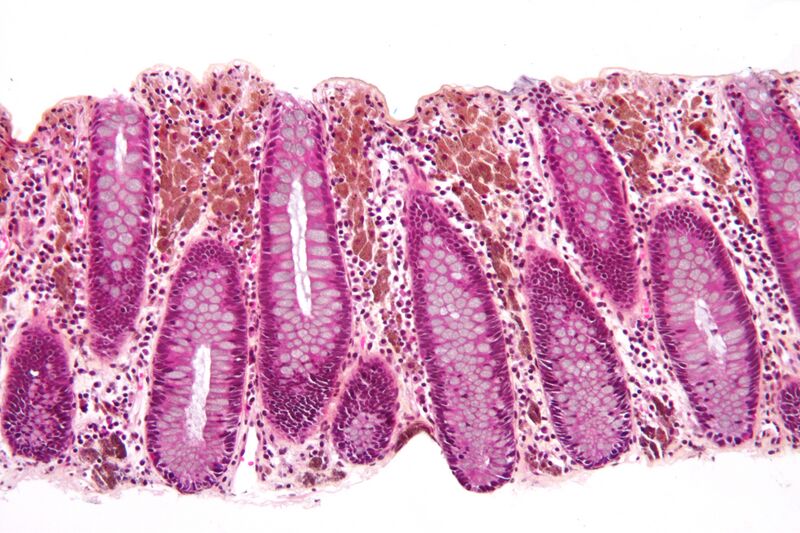
| Description | Intermediate magnification micrograph of melanosis coli (MC), also pseudomelanosis coli (PMC). Colonic biopsy. HPS stain.
Melanosis coli is a misnomer, as the pigmentation is due to lipofuscin-laden macrophages - not melanin pigment. Pseudomelanosis coli is a more appropriate descriptor, but not in common usage. The differential diagnosis of brown pigmentation of the colon is: Pseudomelanosis coli. Hemosiderin-laden macrophages (old haemorrhage). Melanin (rare). PMC is predominantly seen in the cecum and right colon (ascending colon) and associated with anthracene laxative use such as senna (Senokot). Related images
Low mag.
Intermed. mag.
High mag.
High mag.
|
| Source | Wikimedia Commons file page |
| Author | Nephron |
| Permission | See original Commons license details. |
Licensing[edit]
Creative Commons Attribution-ShareAlike 3.0 Unported (CC BY-SA 3.0)
This file is licensed under the Creative Commons Attribution-ShareAlike 3.0 license.
Official license: CC BY-SA 3.0
License page: CC BY-SA 3.0
Original attribution and file history: Wikimedia Commons
File history
Click on a date/time to view the file as it appeared at that time.
| Date/Time | Thumbnail | Dimensions | User | Comment | |
|---|---|---|---|---|---|
| current | 22:27, 8 June 2026 | | 4,272 × 2,848 (3.6 MB) | Maintenance script (talk | contribs) | == Summary == Importing file |
You cannot overwrite this file.
File usage
The following file is a duplicate of this file (more details):
- File:Melanosis coli intermed mag.jpg from Wikimedia Commons
The following page uses this file:
