File:Mesothelioma cytology 1.jpg
Original file (4,272 × 2,848 pixels, file size: 1.45 MB, MIME type: image/jpeg)
Summary[edit]
| Summary | |
|---|---|
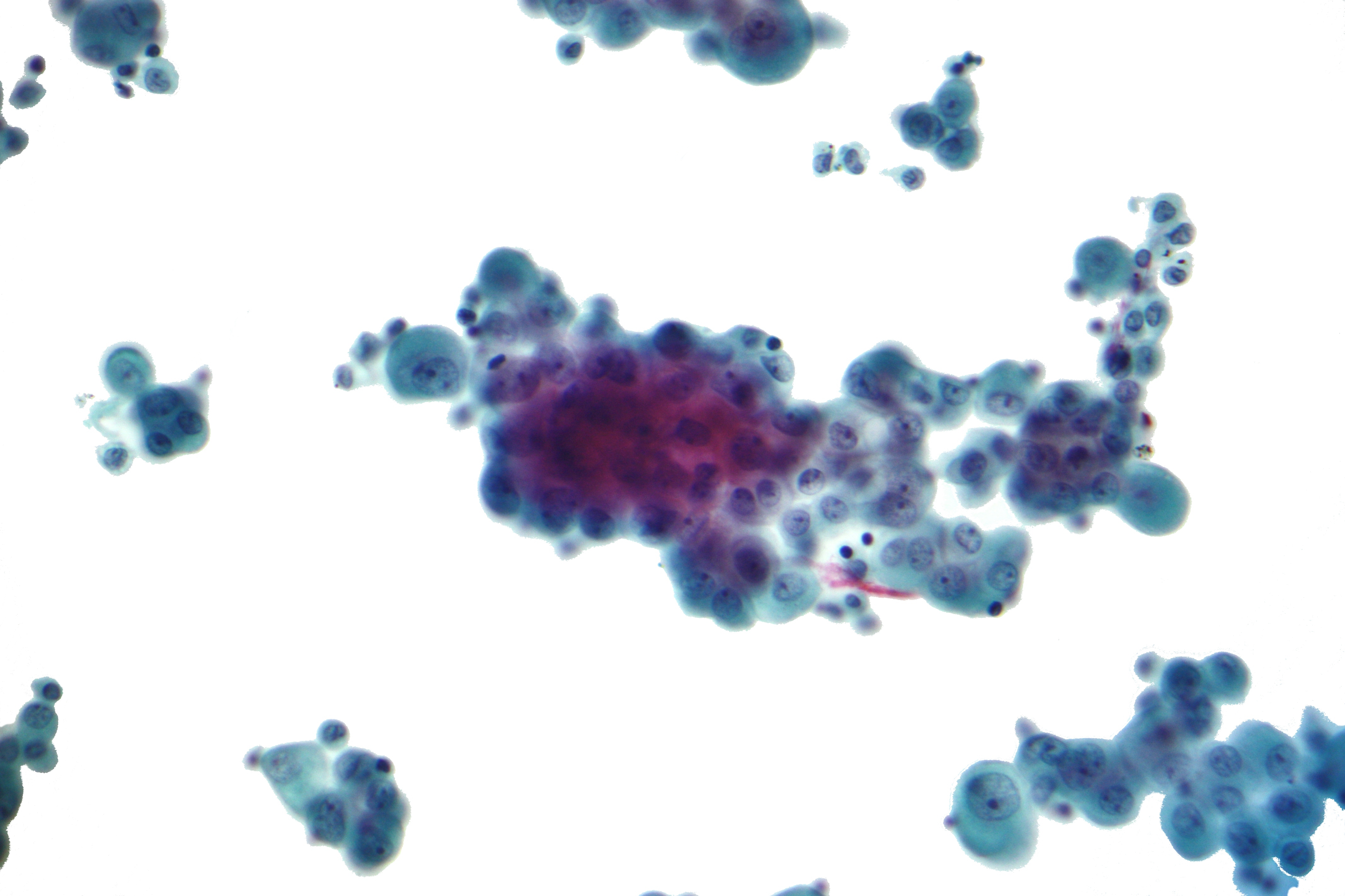
| Description | Micrograph of malignant mesothelioma, also mesothelioma. Cytopathology specimen - pleural fluid.
The image shows the features of mesothelioma: Nuclear membrane irregularies. 3-D clusters of more than 10 cells with "knobby" borders. Large NC ratio (focal). Occasional gigantic cells. Macronucleoli. Multiple nucleoli. Related images
|
| Source | Wikimedia Commons file page |
| Author | Nephron |
| Permission | See original Commons license details. |
Licensing[edit]
Creative Commons Attribution-ShareAlike 3.0 Unported (CC BY-SA 3.0)
This file is licensed under the Creative Commons Attribution-ShareAlike 3.0 license.
Official license: CC BY-SA 3.0
License page: CC BY-SA 3.0
Original attribution and file history: Wikimedia Commons
File history
Click on a date/time to view the file as it appeared at that time.
| Date/Time | Thumbnail | Dimensions | User | Comment | |
|---|---|---|---|---|---|
| current | 03:21, 5 June 2026 | | 4,272 × 2,848 (1.45 MB) | Maintenance script (talk | contribs) | == Summary == Importing file |
You cannot overwrite this file.
File usage
The following file is a duplicate of this file (more details):
- File:Mesothelioma cytology 1.jpg from Wikimedia Commons
The following 2 pages use this file:
