File:Molarsindevelopment11-24-05.jpg
From WikiMD's WELLNESSPEDIA
Size of this preview: 800 × 532 pixels. Other resolution: 1,844 × 1,227 pixels.
Original file (1,844 × 1,227 pixels, file size: 532 KB, MIME type: image/jpeg)
Summary[edit]
| Summary | |
|---|---|
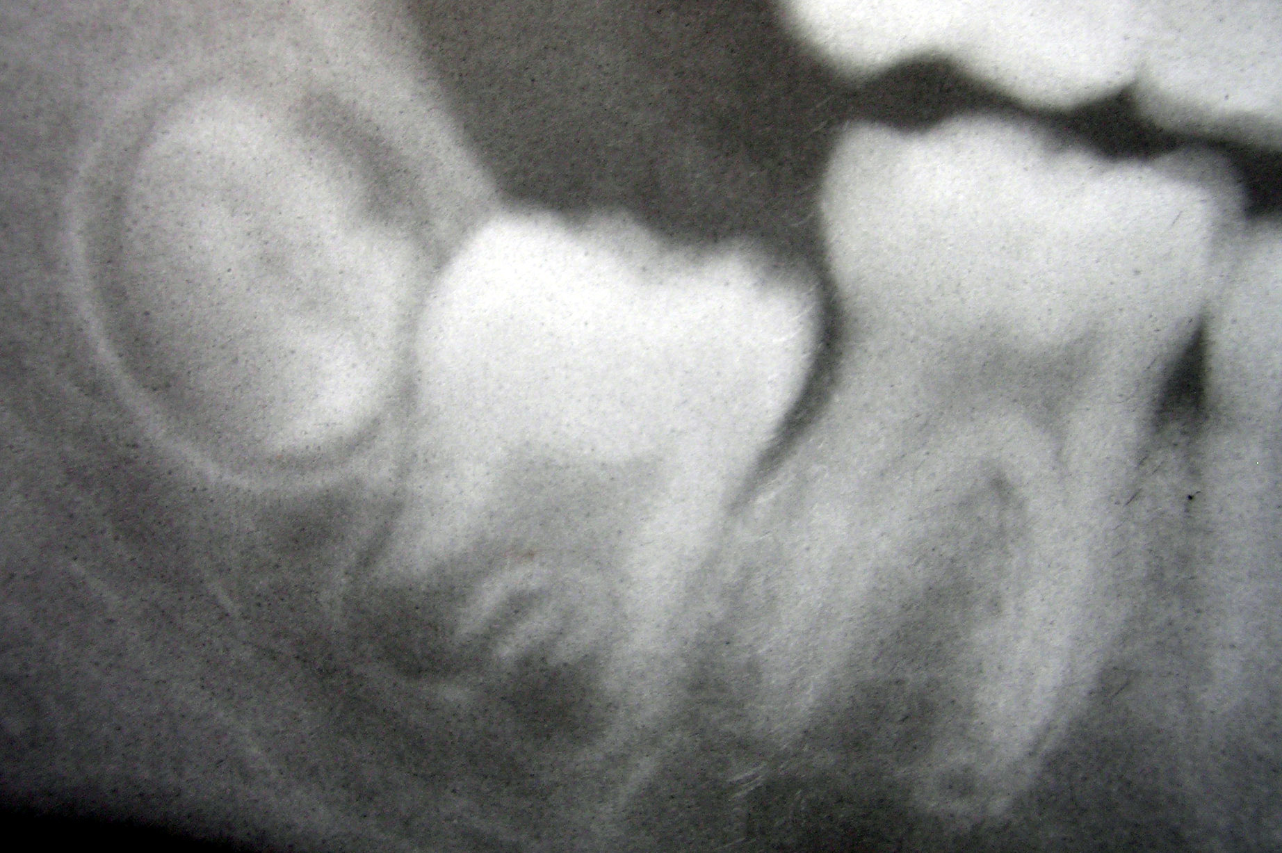
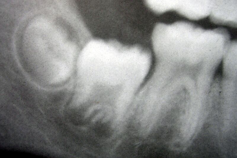
| Description | Radiografía del tercer, segundo y primer molar de la mandíbula, de izquierda a derecha, en distintos estadios de desarrollo. |
| Source | Wikimedia Commons file page |
| Author | No machine-readable author provided. Dozenist assumed (based on copyright claims). |
| Permission | See original Commons license details. |
Licensing[edit]
Creative Commons Attribution-ShareAlike 3.0 Unported (CC BY-SA 3.0)
This file is licensed under the Creative Commons Attribution-ShareAlike 3.0 license.
Official license: CC BY-SA 3.0
Original attribution and file history: Wikimedia Commons
File history
Click on a date/time to view the file as it appeared at that time.
| Date/Time | Thumbnail | Dimensions | User | Comment | |
|---|---|---|---|---|---|
| current | 12:48, 29 May 2026 | | 1,844 × 1,227 (532 KB) | Maintenance script (talk | contribs) | == Summary == Importing file |
You cannot overwrite this file.
File usage
The following file is a duplicate of this file (more details):
- File:Molarsindevelopment11-24-05.jpg from Wikimedia Commons
The following 3 pages use this file:
