File:Mpyv vp1 pentamer vp2 1sie 1cn3.png
From WikiMD's WELLNESSPEDIA
Size of this preview: 675 × 600 pixels. Other resolution: 1,260 × 1,120 pixels.
Original file (1,260 × 1,120 pixels, file size: 702 KB, MIME type: image/png)
Summary[edit]
| Summary | |
|---|---|
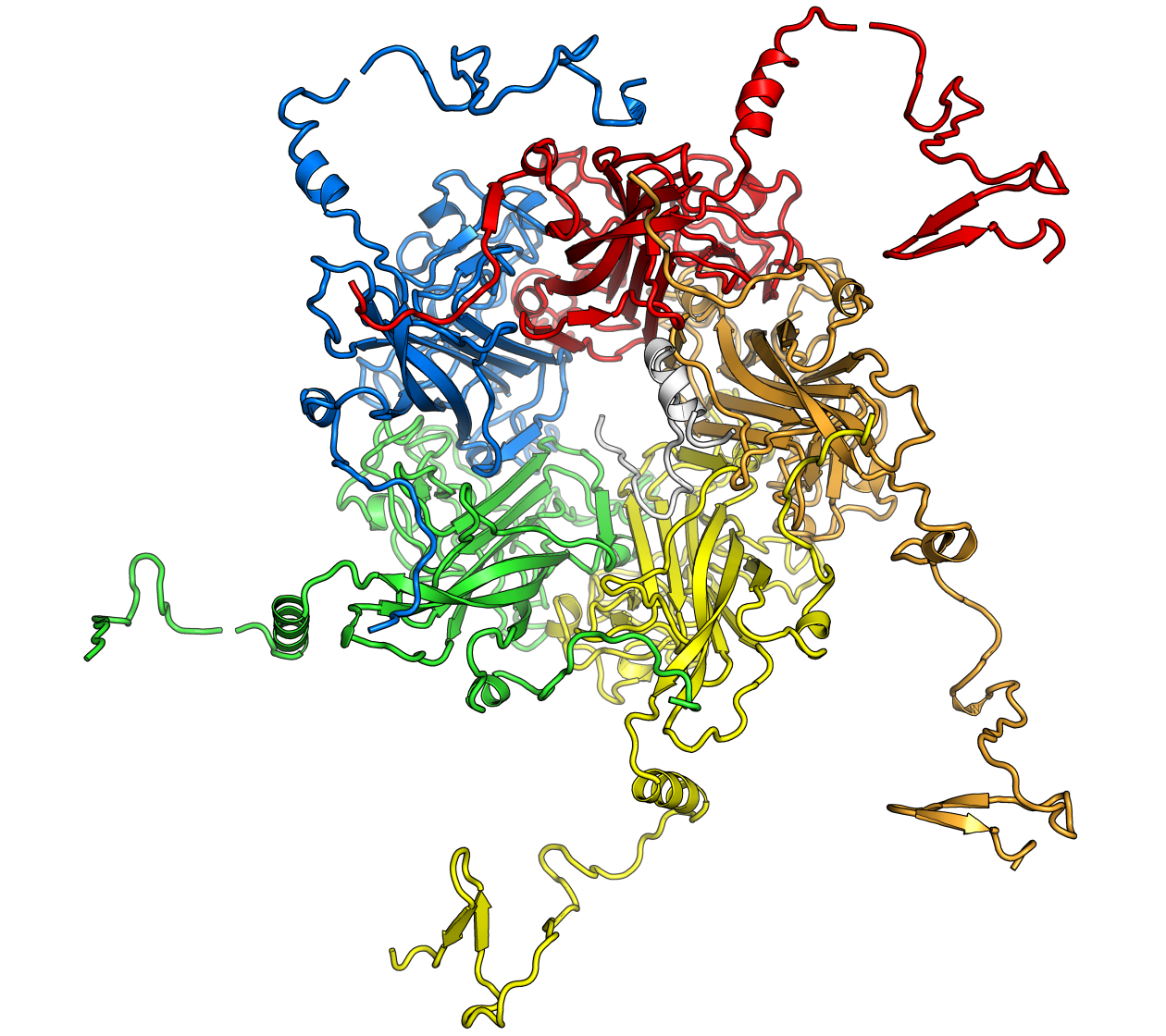
| Description | The structure of an individual pentamer of the murine polyomavirus VP1 protein. Each monomer is colored differently. The conformationally flexible C-terminal arms are shown here in conformations compatible with binding to neighboring molecules. Superposed is a fragment of the polyomavirus VP2 protein (white), which binds to a pentamer oriented toward the central cavity. VP1 is from PDB ID 1SIE; VP2 is from PDB ID 1CN3. |
| Source | Wikimedia Commons file page |
| Author | Opabinia regalis |
| Permission | See original Commons license details. |
Licensing[edit]
Creative Commons Attribution-ShareAlike 3.0 Unported (CC BY-SA 3.0)
This file is licensed under the Creative Commons Attribution-ShareAlike 3.0 license.
Official license: CC BY-SA 3.0
License page: CC BY-SA 3.0
Original attribution and file history: Wikimedia Commons
File history
Click on a date/time to view the file as it appeared at that time.
| Date/Time | Thumbnail | Dimensions | User | Comment | |
|---|---|---|---|---|---|
| current | 22:33, 8 June 2026 | | 1,260 × 1,120 (702 KB) | Maintenance script (talk | contribs) | == Summary == Importing file |
You cannot overwrite this file.
File usage
The following page uses this file:
