File:Multiple myeloma skull CT arrows.PNG
From WikiMD's WELLNESSPEDIA
No higher resolution available.
Multiple_myeloma_skull_CT_arrows.PNG (718 × 324 pixels, file size: 224 KB, MIME type: image/png)
Summary[edit]
| Summary | |
|---|---|
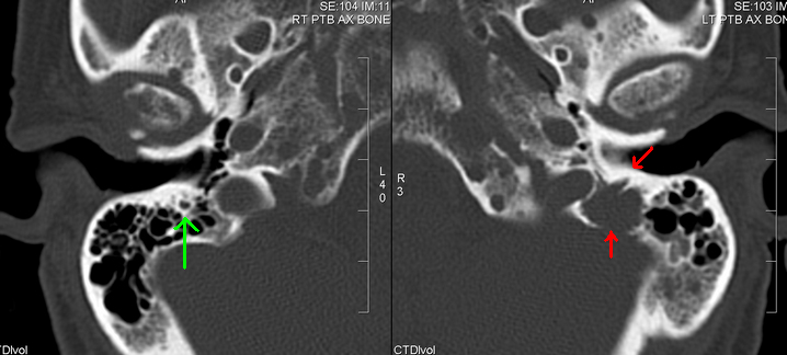
| Description | This 59 year-old patient presented with a left facial droop and a known history of multiple myeloma. A CT brain was performed looking for a cerebral cause. The brain appeared normal. Close inspection revealed a lytic lesion in the left temporal bone, and focused reconstructions of the petrous temporal bones confirmed a lytic lesion involving the mastoid segment of the facial nerve canal. Red arrows: lesion; green arrow: normal contralateral facial nerve canal. The lytic lesion was one of many in the skull and is consistent with a myeloma deposit. |
| Source | Wikimedia Commons file page |
| Author | dr Laughlin Dawes |
| Permission | See original Commons license details. |
Licensing[edit]
Creative Commons Attribution 3.0 Unported (CC BY 3.0)
This file is licensed under the Creative Commons Attribution 3.0 license.
Official license: CC BY 3.0
License page: CC BY 3.0
Original attribution and file history: Wikimedia Commons
File history
Click on a date/time to view the file as it appeared at that time.
| Date/Time | Thumbnail | Dimensions | User | Comment | |
|---|---|---|---|---|---|
| current | 22:25, 8 June 2026 | | 718 × 324 (224 KB) | Maintenance script (talk | contribs) | == Summary == Importing file |
You cannot overwrite this file.
File usage
The following page uses this file:
