File:Nodular fasciitis - intermed mag.jpg
Original file (4,272 × 2,848 pixels, file size: 6.16 MB, MIME type: image/jpeg)
Summary[edit]
| Summary | |
|---|---|
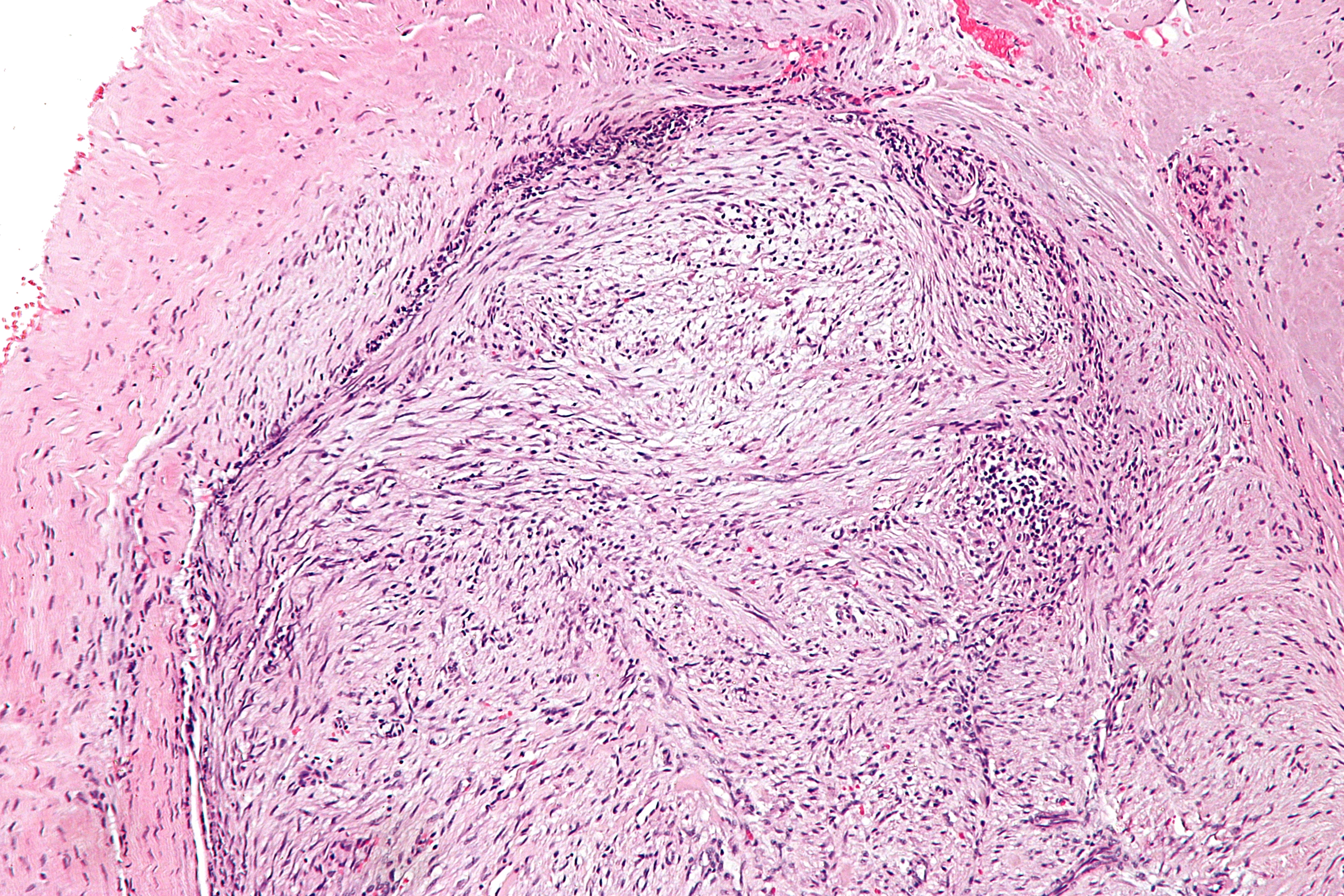
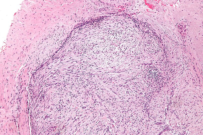
| Description | Intermediate magnification micrograph of nodular fasciitis. H&E stain.
The images show: Tissue culture-like pattern - haphazardly arranged spindle cells. Red blood cell extravasation. Inflammatory cells (predominantly lymphocytes). Other features - not seen: Thick keloid-like collagen. Giant cells. Related images
Low mag.
Intermed. mag.
High mag.
Very high mag.
Intermed. mag.
High mag.
|
| Source | Wikimedia Commons file page |
| Author | Nephron |
| Permission | See original Commons license details. |
Licensing[edit]
Creative Commons Attribution-ShareAlike 3.0 Unported (CC BY-SA 3.0)
This file is licensed under the Creative Commons Attribution-ShareAlike 3.0 license.
Official license: CC BY-SA 3.0
License page: CC BY-SA 3.0
Original attribution and file history: Wikimedia Commons
File history
Click on a date/time to view the file as it appeared at that time.
| Date/Time | Thumbnail | Dimensions | User | Comment | |
|---|---|---|---|---|---|
| current | 22:28, 8 June 2026 | | 4,272 × 2,848 (6.16 MB) | Maintenance script (talk | contribs) | == Summary == Importing file |
You cannot overwrite this file.
File usage
The following file is a duplicate of this file (more details):
- File:Nodular fasciitis - intermed mag.jpg from Wikimedia Commons
The following page uses this file:
