File:Nondisjunction Diagrams.svg
From WikiMD's WELLNESSPEDIA

Size of this PNG preview of this SVG file: 512 × 191 pixels. Other resolution: 2,560 × 955 pixels.
Original file (SVG file, nominally 512 × 191 pixels, file size: 89 KB)
Summary[edit]
| Summary | |
|---|---|
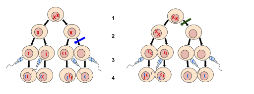
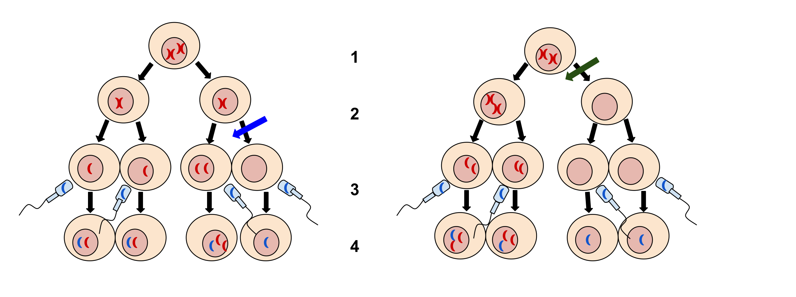
| Description | 1. Meiosis I 2. Meiosis II 3. Fertilization 4. Zygote
The left image at the blue arrow is nondisjunction taking place during meiosis II. The right image at the green arrow is nondisjunction taking place during meiosis I.
|
| Source | Wikimedia Commons file page |
| Author | Tweety207 |
| Permission | See original Commons license details. |
Licensing[edit]
Creative Commons Attribution-ShareAlike 3.0 Unported (CC BY-SA 3.0)
This file is licensed under the Creative Commons Attribution-ShareAlike 3.0 license.
Official license: CC BY-SA 3.0
License page: CC BY-SA 3.0
Original attribution and file history: Wikimedia Commons
File history
Click on a date/time to view the file as it appeared at that time.
| Date/Time | Thumbnail | Dimensions | User | Comment | |
|---|---|---|---|---|---|
| current | 03:30, 5 June 2026 | 512 × 191 (89 KB) | Maintenance script (talk | contribs) | == Summary == Importing file |
You cannot overwrite this file.
File usage
The following 2 pages use this file:
