File:OCD WalterReed MRI-Sagital-T1.jpeg
From WikiMD's WELLNESSPEDIA
Size of this preview: 579 × 600 pixels. Other resolution: 945 × 979 pixels.
Original file (945 × 979 pixels, file size: 353 KB, MIME type: image/jpeg)
Summary[edit]
| Summary | |
|---|---|
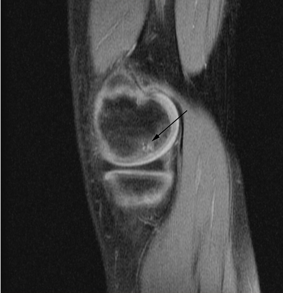
| Description | "Sagittal and coronal T1 and T2 images demonstrate linear low T1, high T2 signal at the articular surfaces of the lateral aspects of the medial femoral condyles bilaterally, corresponding to the radiographs, confirming the presence of bilateral osteochondritis dissecans, with diffuse increase in T2 signal at the medial femoral condyles, indicating marrow edema." From the case of a 9-year-old buy with bilateral knee pain. |
| Source | Wikimedia Commons file page |
| Author | Pil Kang |
| Permission | See original Commons license details. |
Licensing[edit]
Public Domain
This file is in the public domain and may be used without restriction.
Please see the linked source page for the original file history, attribution information, and licensing details.
Original attribution and file history: Wikimedia Commons
File history
Click on a date/time to view the file as it appeared at that time.
| Date/Time | Thumbnail | Dimensions | User | Comment | |
|---|---|---|---|---|---|
| current | 01:21, 2 June 2026 | | 945 × 979 (353 KB) | Maintenance script (talk | contribs) | == Summary == Importing file |
You cannot overwrite this file.
File usage
The following 2 pages use this file:
