File:Pap stain of adenocarcinoma in peritoneal fluid.png
From WikiMD's WELLNESSPEDIA
Size of this preview: 707 × 600 pixels. Other resolution: 1,149 × 975 pixels.
Original file (1,149 × 975 pixels, file size: 856 KB, MIME type: image/png)
Summary[edit]
| Summary | |
|---|---|
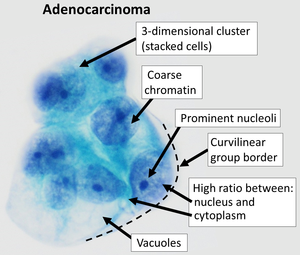
| Description | Cytopathology of peritoneal fluid in a case of peritoneal carcinomatosis (Pap stain), showing typical features of adenocarcinoma. Vacuoles are more prominent in mucinous tumors, but can be seen in serous tumors as well. |
| Source | Wikimedia Commons file page |
| Author | Mikael Häggström, M.D. Author info - Reusing images- Conflicts of interest: None Mikael Häggström, M.D.Consent note: Consent from the patient or patient's relatives is regarded as redundant, because of absence of identifiable features (List of HIPAA identifiers) in the media and case information (See also HIPAA case reports guidance). |
| Permission | See original Commons license details. |
Licensing[edit]
License: CC0
License page: CC0
Original attribution and file history: Wikimedia Commons
File history
Click on a date/time to view the file as it appeared at that time.
| Date/Time | Thumbnail | Dimensions | User | Comment | |
|---|---|---|---|---|---|
| current | 03:21, 5 June 2026 | | 1,149 × 975 (856 KB) | Maintenance script (talk | contribs) | == Summary == Importing file |
You cannot overwrite this file.
File usage
The following file is a duplicate of this file (more details):
- File:Pap stain of adenocarcinoma in peritoneal fluid.png from Wikimedia Commons
The following 2 pages use this file:
