File:Periapical radiopaque-hyperdense jaw lesions 2.jpg
From WikiMD's WELLNESSPEDIA
Size of this preview: 411 × 600 pixels. Other resolution: 900 × 1,313 pixels.
Original file (900 × 1,313 pixels, file size: 143 KB, MIME type: image/jpeg)
Summary[edit]
| Summary | |
|---|---|
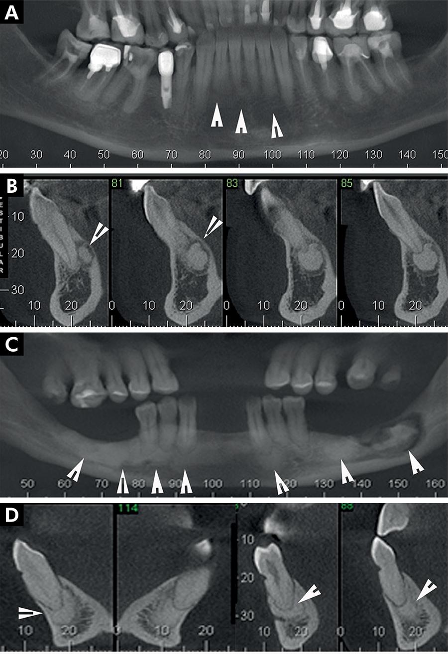
| Description | Periapical cemento-osseous dysplasia (periapical COD): Panoramic images showing hyperdense round lesions in the periapical region of a mandibular anterior sound tooth (arrowhead) (A). Cross-sectional images showing periapical COD lesions markedly separated from the adjacent normal bone and also separated from the root of the anterior tooth (arrowhead) (B). Florid COD: In CBCT images, hyperdense multiple lesions are observed that involve the mandible bilaterally (arrow head) and bone sequestration in left mandible (arrow) (C). In (D), hyperdense elliptical lesions are seen clearly separated from the root of a mandibular tooth (arrowhead). |
| Source | Wikimedia Commons file page |
| Author | SILVA, Brunno Santos Freitas (2017). "Differential diagnosis and clinical management of periapical radiopaque/hyperdense jaw lesions". Brazilian Oral Research 31 (0). FapUNIFESP (SciELO). DOI:10.1590/1807-3107bor-2017.vol31.0052. ISSN 1806-8324. |
| Permission | See original Commons license details. |
Licensing[edit]
Creative Commons Attribution 4.0 International (CC BY 4.0)
This file is licensed under the Creative Commons Attribution 4.0 International license.
You may share and adapt the material provided appropriate attribution is given.
Official license: CC BY 4.0
License page: CC BY 4.0
Original attribution and file history: Wikimedia Commons
File history
Click on a date/time to view the file as it appeared at that time.
| Date/Time | Thumbnail | Dimensions | User | Comment | |
|---|---|---|---|---|---|
| current | 03:25, 5 June 2026 | | 900 × 1,313 (143 KB) | Maintenance script (talk | contribs) | == Summary == Importing file |
You cannot overwrite this file.
File usage
The following file is a duplicate of this file (more details):
- File:Periapical radiopaque-hyperdense jaw lesions 2.jpg from Wikimedia Commons
The following 2 pages use this file:
