File:Polymicrogyria arrows.JPG
From WikiMD's WELLNESSPEDIA
No higher resolution available.
Polymicrogyria_arrows.JPG (610 × 598 pixels, file size: 80 KB, MIME type: image/jpeg)
Summary[edit]
| Summary | |
|---|---|
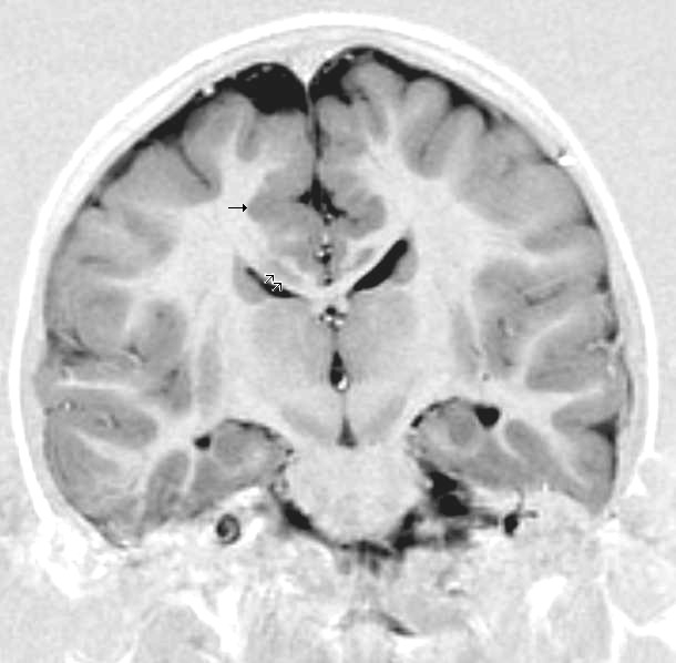
| Description | This child presented with seizures. The coronal true inversion recovery sequence shows thickened and disordered cortex in superior frontal and cingulate gyri bilaterally (arrow). There are small convolutions visible at the corticomedullary junction. The appearance is that of cortical dysplasia, with polymicrogyria more likely than pachygyria due to the small convolutions visible. There are also small foci of grey matter signal in the corpus callosum, deep to the dysplastic cortex (double arrows). These probably represent areas of grey matter heterotopia. |
| Source | Wikimedia Commons file page |
| Author | Dr Laughlin Dawes |
| Permission | See original Commons license details. |
Licensing[edit]
Creative Commons Attribution 3.0 Unported (CC BY 3.0)
This file is licensed under the Creative Commons Attribution 3.0 license.
Official license: CC BY 3.0
License page: CC BY 3.0
Original attribution and file history: Wikimedia Commons
File history
Click on a date/time to view the file as it appeared at that time.
| Date/Time | Thumbnail | Dimensions | User | Comment | |
|---|---|---|---|---|---|
| current | 03:35, 5 June 2026 | | 610 × 598 (80 KB) | Maintenance script (talk | contribs) | == Summary == Importing file |
You cannot overwrite this file.
File usage
The following 2 pages use this file:
